SISTEM PENCERNAAN RUMINANSIA Proses Pencernaan Sapi
advertisement
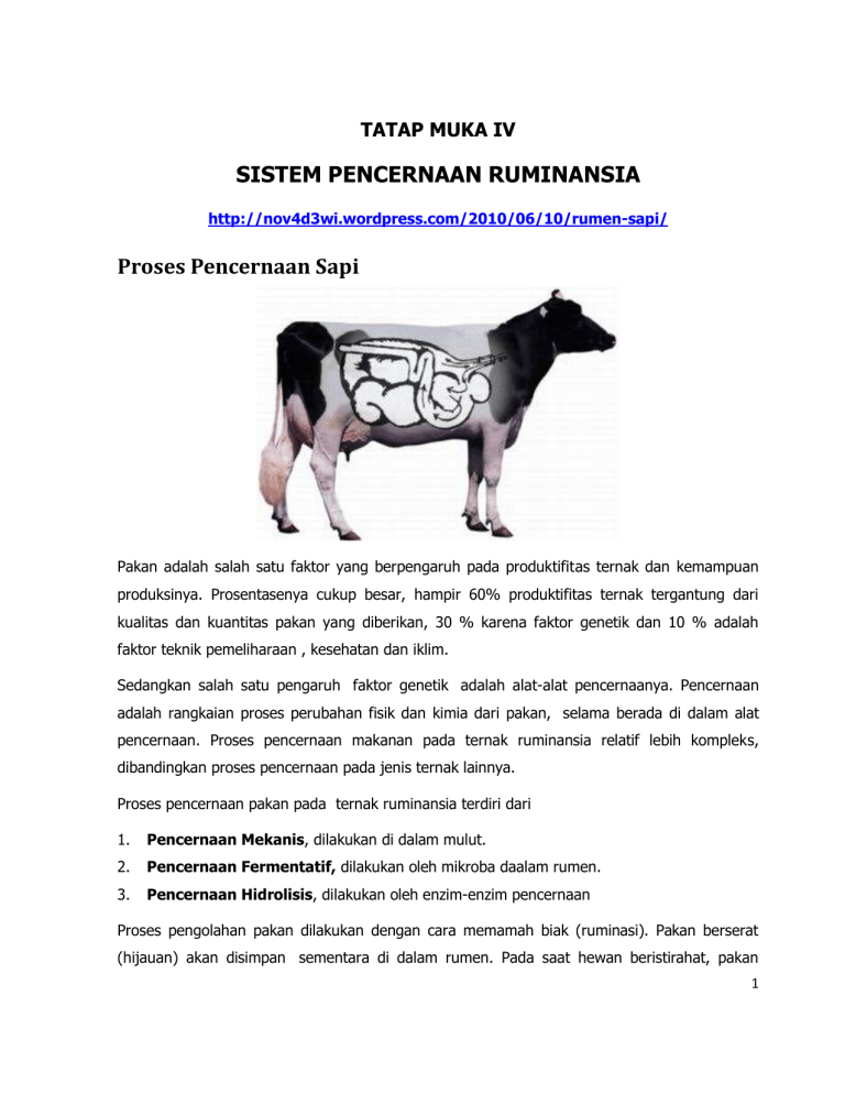
TATAP MUKA IV SISTEM PENCERNAAN RUMINANSIA http://nov4d3wi.wordpress.com/2010/06/10/rumen-sapi/ Proses Pencernaan Sapi Pakan adalah salah satu faktor yang berpengaruh pada produktifitas ternak dan kemampuan produksinya. Prosentasenya cukup besar, hampir 60% produktifitas ternak tergantung dari kualitas dan kuantitas pakan yang diberikan, 30 % karena faktor genetik dan 10 % adalah faktor teknik pemeliharaan , kesehatan dan iklim. Sedangkan salah satu pengaruh faktor genetik adalah alat-alat pencernaanya. Pencernaan adalah rangkaian proses perubahan fisik dan kimia dari pakan, selama berada di dalam alat pencernaan. Proses pencernaan makanan pada ternak ruminansia relatif lebih kompleks, dibandingkan proses pencernaan pada jenis ternak lainnya. Proses pencernaan pakan pada ternak ruminansia terdiri dari 1. Pencernaan Mekanis, dilakukan di dalam mulut. 2. Pencernaan Fermentatif, dilakukan oleh mikroba daalam rumen. 3. Pencernaan Hidrolisis, dilakukan oleh enzim-enzim pencernaan Proses pengolahan pakan dilakukan dengan cara memamah biak (ruminasi). Pakan berserat (hijauan) akan disimpan sementara di dalam rumen. Pada saat hewan beristirahat, pakan 1 akan ditarik kembali ke mulut (proses regurgitasi),untuk dikunyah (proses remastikasi). Selanjutnya pakan akan ditelan (proses redeglutasi)., untuk dicerna oleh enzim-enzim mikroba rumen. Di dalam perut, pakan akan diolah di 4 kompartemen perut, yaitu : 1. Retikulum (perut jala). 2. Rumen (perut beludru). 3. Omasum (perut buku,tersusun dari +/- 100 lipatan ). 4. Abomasum (perut/lambung sejati,karena baik anatomis maupun fisiologisnya sama dengan lambung non-ruminansia). Alat pencernaan sapi ini , berkembang dalam 3 fase sesuai dengan umur sapi yaitu : Fase Non Ruminansi. Fase ini terjadi pada pedet yang baru lahir. Volume retikulo-rumen pada pedet yang baru lahir hanya sekitar 30% dari total kapasitas total perut dan rumennya masih belum berfungsi. Oleh sebab itu, pada fase ini Nutrisi didapat hanya dari susu yang berasal dari induknya. Proses pengolahanya pun langsung ke omasum (tanpa melewati rumen), melalui suatu saluran yang disebut esophagial groove. Saluran ini menghubungkan esophagus dan reticular omasal orifice. Fase Transisi. Fase ini terjadi pada pedet yang telah berusia 2 minggu. Pada usia ini pedet akan mulai belajar memakan pakan kasar (hijauan). Secara bertahap rumen juga berkembang, lebih cepat dari pada kompartemen perut yang lain. Pada fase ini pula mikroba mulai dan rumen mulai berfungsi sebagai tempat fermentasi karbohidrat. Fase Ruminansia. Fase ini terjadi pada pedet yang telah berumur 6 minggu. Alat pencernaan mulai berkembang menuju kesempurnaan, hingga komposisi rumen mencapai 81%, retikulum 3%, omasum 7%, dan abomasum 9% dari volume total perut. 2 Proses Pencernaan di Mulut Setelah masuk kedalam mulut sapi, pakan akan diolah secara mekanis (dihancurkan) oleh gigi. Kemudian pakan akan bercampur dengan saliva (air liur), yang disekresikan oleh 3 pasang glandula saliva, yaitu glandula parotid yang terletak di depan telinga, glandula submandibularis (sumbaxillaris) yang terletak pada rahang bawah, dan glandula sublingualis yang terletak dibawah lidah. Kandungan Saliva terdiri dari air sebanyak 99% airdan 1% sisanya terdiri atas mucin, garamgaram anorganik, dan lisozim kompleks. Saliva pada sapi juga mengandung urea, fosfor (P), dan natrium (Na) yang dapat dimanfaatkan oleh mikroba rumen. Tetapi Saliva pada sapi tidak mengandung enzim -amilase yang dapat membantu proses pencernaan. Fungsi saliva adalah untuk : 1. Membasahi pakan agar mudah ditelan 2. Menjaga pH rumen agar tidak naik atau turun terlalu tajam, hal ini terjadi karena saliva memiliki sifat buffer (penyangga) dari bikarbinat yang terkandung didalamnya. 3 Pencernaan pada ternak dimulai dari mulut yang di dalamnya terdapat gigi geligi, yaitu : 1. Gigi seri (Insisivus) memiliki bentuk untuk menjepit makanan berupa tetumbuhan seperli rumput. 2. Geraham belakang (Molare) memiliki bentuk datar dan lobar. 3. Rahang dapat bergerak menyamping untuk menggiling makanan. Pola sistem pencernaan pada hewan umumnya sama dengan manusia, yaitu terdiri atas mulut, faring, esofagus, lambung, dan usus. Namun demikian, struktur alat pencernaan kadangkadang berbeda antara hewan yang satu dengan hewan yang lain. Sapi, misalnya, mempunyai susunan gigi sebagai berikut: 3 3 0 0 0 0 0 0 Rahang atas M P C I I C P M Jenis gigi 3 3 0 4 4 0 3 3 Rahang bawah I = insisivus = gigi seri C = kaninus = gigi taring P = premolar = geraham depan M = molar = geraham belakang Berdasarkan susunan gigi di atas, terlihat bahwa sapi (hewan memamah biak) tidak mempunyai gigi seri bagian atas dan gigi taring, tetapi memiliki gigi geraham lebih banyak dibandingkan dengan manusia sesuai dengan fungsinya untuk mengunyah makanan berserat, yaitu penyusun dinding sel tumbuhan yang terdiri atas 50% selulosa. Jika dibandingkan dengan kuda, faring pada sapi lebih pendek. Esofagus (kerongkongan) pada sapi sangat pendek dan lebar serta lebih mampu berdilatasi (mernbesar). Esofagus berdinding tipis dan panjangnya bervariasi diperkirakan sekitar 5 cm. Lambung sapi sangat besar, diperkirakan sekitar 3/4 dart isi rongga perut. Lambung mempunyai peranan penting untuk menyimpan makanan sementara yang akan dimamah kembali (kedua kah). Selain itu, pada lambung juga terjadi proses pembusukan dan peragian. 4 Lambung ruminansia terdiri atas 4 bagian, yaitu rumen, retikulum, omasum, dan abomasum dengan ukuran yang bervariasi sesuai dengan umur dan makanan alamiahnya. Kapasitas rumen 80%, retikulum 5%, omasum 7-8%, dan abomasum 7-8%. Pembagian ini terlihat dari bentuk gentingan pada saat otot sfinkter berkontraksi. Makanan dari kerongkongan akan masuk rumen yang berfungsi sebagai gudang sementara bagi makanan yang tertelan. Di rumen terjadi pencernaan protein, polisakarida, dan fermentasi selulosa oleh enzim selulase yang dihasilkan oleh bakteri dan jenis protozoa tertentu. Dari rumen, makanan akan diteruskan ke retikulum dan di tempat ini makanan akan dibentuk menjadi gumpalan-gumpalan yang masih kasar (disebut bolus). Bolus akan dimuntahkan kembali ke mulut untuk dimamah kedua kali. Dari mulut makanan akan ditelan kembali untuk diteruskan ke ornasum. Pada omasum terdapat kelenjar yang memproduksi enzim yang akan bercampur dengan bolus. Akhirnya bolus akan diteruskan ke abomasum, yaitu perut yang sebenarnya dan di tempat ini masih terjadi proses pencernaan bolus secara kimiawi oleh enzim. Selulase yang dihasilkan oleh mikroba (bakteri dan protozoa) akan merombak selulosa menjadi asam lemak. Akan tetapi, bakteri tidak tahan hidup di abomasum karena pH yang sangat rendah, akibatnya bakteri ini akan mati, namun dapat dicernakan untuk menjadi sumber protein bagi hewan pemamah biak. Dengan demikian, hewan ini tidak memerlukan asam amino esensial seperti pada manusia. Hewan seperti kuda, kelinci, dan marmut tidak mempunyai struktur lambung seperti pada sapi untuk fermentasi seluIosa. Proses fermentasi atau pembusukan yang dilaksanakan oleh bakteri terjadi pada sekum yang banyak mengandung bakteri. Proses fermentasi pada sekum tidak seefektif fermentasi yang terjadi di lambung. Akibatnya kotoran kuda, kelinci, dan marmut lebih kasar karena proses pencernaan selulosa hanya terjadi satu kali, yakni pada sekum. Sedangkan pada sapi proses pencernaan terjadi dua kali, yakni pada lambung dan sekum yang kedua-duanya dilakukan oleh bakteri dan protozoa tertentu. Pada kelinci dan marmut, kotoran yang telah keluar tubuh seringkali dimakan kembali. Kotoran yang belum tercerna tadi masih mengandung banyak zat makanan, yang akan dicernakan lagi oleh kelinci. 5 Sekum pada pemakan tumbuh-tumbuhan lebih besar dibandingkan dengan sekum karnivora. Hal itu disebabkan karena makanan herbivora bervolume besar dan proses pencernaannya berat, sedangkan pada karnivora volume makanan kecil dan pencernaan berlangsung dengan cepat. Usus pada sapi sangat panjang, usus halusnya bisa mencapai 40 meter. Hal itu dipengaruhi oleh makanannya yang sebagian besar terdiri dari serat (selulosa). Enzim selulase yang dihasilkan oleh bakteri ini tidak hanya berfungsi untuk mencerna selulosa menjadi asam lemak, tetapi juga dapat menghasilkan bio gas yang berupa CH4 yang dapat digunakan sebagai sumber energi alternatif. Tidak tertutup kemungkinan bakteri yang ada di sekum akan keluar dari tubuh organisme bersama feses, sehingga di dalam feses (tinja) hewan yang mengandung bahan organik akan diuraikan dan dapat melepaskan gas CH4 (gas bio). Sumber: http://bebas.vlsm.org/v12/sponsor/Sponsor Pendamping/Praweda/Biologi/0066% 20Bio%202-5d.htm Sistem Pencernaan Ruminansia Pencernaan adalah rangkaian proses perubahan fisik dan kimia yang dialami bahan makanan selama berada di dalam alat pencernaan. Proses pencernaan makanan pada ternak ruminansia relatif lebih kompleks dibandingkan proses pencernaan pada jenis ternak lainnya. Perut ternak ruminansia dibagi menjadi 4 bagian, yaitu retikulum (perut jala), rumen (perut beludru), omasum (perut bulu), dan abomasum (perut sejati). Dalam studi fisiologi ternak ruminasia, rumen dan retikulum sering dipandang sebagai organ tunggal dengan sebutan retikulo-rumen. Omasum disebut sebagai perut buku karena tersusun dari lipatan sebanyak sekitar 100 lembar. Fungsi omasum belum terungkap dengan jelas, tetapi pada organ tersebut terjadi penyerapan air, amonia, asam lemak terbang dan elektrolit. Pada organ ini dilaporkan juga menghasilkan amonia dan mungkin asam lemak terbang (Frances dan Siddon, 1993). Termasuk organ pencernaan bagian belakang lambung adalah sekum, kolon dan rektum. Pada pencernaan bagian belakang tersebut juga terjadi aktivitas fermentasi. Namun belum banyak informasi yang terungkap tentang peranan fermentasi pada organ tersebut, yang terletak setelah organ penyerapan utama. Proses pencernaan pada ternak ruminansia dapat terjadi 6 secara mekanis di mulut, fermentatif oleh mikroba rumen dan secara hidrolis oleh enzim-enzim pencernaan. Pada sistem pencernaan ternak ruminasia terdapat suatu proses yangdisebut memamah biak (ruminasi). Pakan berserat (hijauan) yang dimakanditahan untuk sementara di dalam rumen. Pada saat hewan beristirahat, pakanyang telah berada dalam rumen dikembalikan ke mulut (proses regurgitasi),untuk dikunyah kembali (proses remastikasi), kemudian pakan ditelan kembali(proses redeglutasi). Selanjutnya pakan tersebut dicerna lagi oleh enzim-enzimmikroba rumen. Kontraksi retikulorumen yang terkoordinasi dalam rangkaianproses tersebut bermanfaat pula untuk pengadukan digesta inokulasi danpenyerapan nutrien. Selain itu kontraksi retikulorumen juga bermanfaat untukpergerakan digesta meninggalkan retikulorumen melalui retikulo-omasal orifice(Tilman et al. 1982). Di dalam rumen terdapat populasi mikroba yang cukup banyak jumlahnya.Mikroba rumen dapat dibagi dalam tiga grup utama yaitu bakteri, protozoa danfungi (Czerkawski, 1986). Kehadiran fungi di dalam rumen diakui sangatbermanfaat bagi pencernaan pakan serat, karena dia membentuk koloni padajaringan selulosa pakan. Rizoid fungi tumbuh jauh menembus dinding seltanaman sehingga pakan lebih terbuka untuk dicerna oleh enzim bakteri rumen. Bakteri rumen dapat diklasifikasikan berdasarkan substrat utama yangdigunakan, karena sulit mengklasifikasikan berdasarkan morfologinya.Kebalikannya protozoa diklasifikasikan berdasarkan morfologinya sebab mudahdilihat berdasarkan penyebaran silianya. Beberapa jenis bakteri yang dilaporkanoleh Hungate (1966) adalah : (a) bakteri pencerna selulosa (Bakteroidessuccinogenes, Ruminococcus flavafaciens, Ruminococcus albus, Butyrifibriofibrisolvens), (b) bakteri pencerna hemiselulosa (Butyrivibrio fibrisolvens,Bakteroides ruminocola, Ruminococcus sp), (c) bakteri pencerna pati(Bakteroides ammylophilus, Streptococcus bovis, Succinnimonas amylolytica, (d) bakteri pencerna gula (Triponema bryantii, Lactobasilus ruminus), (e) bakteri pencerna protein (Clostridium sporogenus, Bacillus licheniformis). Protozoa rumen diklasifikasikan menurut morfologinya yaitu: Holotrichs yang mempunyai silia hampir diseluruh tubuhnya dan mencerna karbohidrat yangfermentabel, sedangkan Oligotrichs yang mempunyai silia sekitar mulutumumnya merombak karbohidrat yang lebih sulit dicerna (Arora, 1989). 7 Rumen merupakan bagian sistem pencernaan pada ternak ruminansia. Pada rumen terjadi pencernaan secara fermentatif dan pencernaan secara hidrolitik. Pencernaan fermentatif memerlukan bantuan mikroba dalam mencerna pakan terutama pakan dengan kandungan selulosa dan hemiselulosa yang tinggi. Sedangkan pencernaan hidrolitik memerlukan bantuan enzim dari sistem pencernaan hewan itu sendiri dalam mencerna pakan. Lambung pada ternak ruminansia dapat dibagi menjadi empat bagian yaitu : 1. Retikulum Retikulum sering disebut sebagai perut jala atau hardware stomach. Retikulum berbatasan langsung dengan rumen, akan tetapi diantara keduanya tidak ada dinding penyekat. Pembatas diantara retikulum dan rumen yaitu hanya berupa lipatan, sehingga partikel pakan menjadi tercampur. 2. Rumen Rumen pada sapi dewasa merupakan bagian yang mempunyai proporsi yang tinggi dibandingkan dengan proporsi bagian lainnya. Rumen terletak di rongga abdominal bagian kiri. Rumen sering disebut juga dengan perut beludru. Hal tersebut dikarenakan pada permukaan rumen terdapat papilla. Pada retikulum dan rumen terjadi pencernaan secara fermentatif, karena pada bagian tersebut terdapat mikroba dengan jumlah bermilyar-milyar. 3. Omasum Omasum sering juga disebut dengan perut buku, karena permukaannya berbuku-buku. Derajat Keasaman (pH) omasum berkisar antara 5,2 sampai 6,5. Antara omasum dan abomasum terdapat lubang yang disebut omaso abomasal orifice. Fungsi omaso abomasal orifice adalah untuk mencegah digesta yang ada di abomasum kembali ke omasum. 4. Abomasum Abomasum sering juga disebut dengan perut sejati. Derajat keasaman (pH) pada abomasum asam yaitu berkisar antara 2 sampai 4,1. Permukaan abomasums dilapisi oleh mukosa yang berfungsi untuk melindungi dinding sel agar tidak tercerna oleh enzim yang dihasilkan oleh abomasum (Priyono, 2009). 8 Rumen Sapi Perut ruminansia terdiri atas retikulum, rumen, omasum dan abomasum. Volume rumen pada ternak sapi dapat mencapai 100 liter atau lebih dan untuk domba berkisar 10 liter. Isi rumen yang merupakan limbah rumah potong hewan bila tidak ditangani dengan baik dapat mencemari lingkungan, sebaliknya isi rumen berpotensi sebagai feed additive. Menurut Gohl (1981) isi rumen dapat mencapai 8-10% dari berat sapi atau kerbau yang dipuasakan sebelum dipotong. Jovanovic dan Cuperlovic (1977) menyatakan mikrobia rumen dapat meningkatkan nilai gizi bahan makanan karena adanya enzim yang dihasilkan oleh mikroba sehingga akan meningkatkan daya cerna. Kualitas isi rumen dipengaruhi oleh jenis makanan, mikrobia rumen dan lamanya makanan dalam rumen. (Barnet dan Nair, 1961). Menurut Jovanovic dan Cuperlvic (1977) kualitas isi rumen tidak begitu bervariasi antara hewan yang dipotong dari berbagai tempat, sebab hewan dipuasakan terlebih dahulu sehingga adanya variasi dari ransum akan teratasi. Di dalam rumen, organisme akan memfermentasi karbohidrat yang spesifik dengan menggunakan enzim untuk mendegradasi substrat sebagai sumber energi. Church (1979) menyatakan bahwa cairan rumen mengandung enzim -amilase, galaktosidase, hemisellulase, sellulase dan xilanaseenzim yang aktif pada xilan dan arabinosa. Enzim proteolitik dan selulolitik juga terdeteksi pada level yang tinggi. Enzim protease terdistribusi berturut-turut sebesar 37,7 ; 22,1 dan 40,2% dan amilase 51,6; 18,2 dan 30,2% pada cairan rumen, partikel pakan dan keseluruhan rumen. Tingkat degradasi perlakuan penambahan enzim cairan rumen 9 dengan aktivitas enzim selulase 2,43 IU/g pada substrat onggok, gaplek, bungkil kelapa, jeram padi dan dedak berturut-turut sebesar 89,9; 85,89; 83,97 ; 72,79 dan 67,81% (Murni,2003). Sumber Pustaka : Priyono, 2009. Rumen Pada Ternak Ruminansia URL http://priyonoscience.blogspot.com/2009/05/rumen-pada-ternak-ruminansia.html) (25/04/2010) : Gohl, BO. 1981. Tropical Feed. Food and Agriculture Organization of The United Nation, Rome. Jovanovic, M. Cuperlvic M. 1977. Nutritive Value Of Rumen Content For Oogastric. Anim Feed Sci and Tech 2:351-360. Murni, S. 2003. Aktivitas Enzim Cairan Rumen Pada Bebrapa Bahan Pakan Dan Pengaruhna Terhadap Performans Broiler Yang Diberi Ransum Berbahan Baku Singkong. Skripsi. Institut Pertanian Bogor. Barnet A.F.G, and Matoba T. Nair, B.M. 1961. Reaction in Rumen. Edward Arnold. Pub. Ltd. London. KANDUNGAN ZAT RUMEN SAPI http://anang-pasi.blogspot.com/2010/07/kandungan-zat-rumen-sapi.html Isi rumen merupakan salah satu limbah rumah potong hewan yang belum dimanfaatkan secara optimal bahkan ada yang dibuang begitu saja sehingga menimbulkan pencemaran lingkungan. Limbah ini sebenarnya sangat potensial bila dimanfaatkan sebagai bahan pakan ternak karena isi rumen disamping merupakan bahan pakan yang belum tercerna juga terdapat organisme rumen yang merupakan sumber vitamin B. Kandungan zat makanan yang terdapat pada isi rumen sapi meliputi: air (8,8%), protein kasar (9,63%), lemak (1,81%), serat kasar (24,60%), BETN (38,40%), Abu (16,76%), kalsium (1,22%) dan posfor (0,29%) dan pada domba meliputi: air (8,28%), protein kasar (14,41%), lemak (3,59%), serat kasar (24,38%), Abu (16,37%), kalsium (0,68%) dan posfor (1,08%) (Suhermiyati, 1984). Widodo (2002) menyatakan zat makanan yang terkandung dalam rumen meliputi protein sebesar 8,86%, lemak 2,60%, serat kasar 28,78%, fosfor 0,55%, abu 18,54% dan air 10,92%. Berdasarkan komposisi zat yang terkandung didalamnya maka isi rumen 10 dalam batas tertentu tidak akan menimbulkan akibat yang merugikan bila dijadikan bahan pencampur ransum berbagai ternak. Di dalam rumen ternak ruminansia (sapi, kerbau, kambing dan domba) terdapat populasi mikroba yang cukup banyak jumlahnya. Cairan rumen mengandung bakteri dan protozoa. Konsentrasi bakteri sekitar 10 pangkat 9 setiap cc isi rumen, sedangkan protozoa bervariasi sekitar 10 pangkat 5 - 10 pangkat 6 setiap cc isi rumen (Tillman, 1991). Beberapa jenis bakteri/mikroba yang terdapat dalam isi rumen adalah (a) bakteri/mikroba lipolitik, (b) bakteri/mikroba pembentuk asam, (c) bakteri/mikroba amilolitik, (d) bakteri/mikroba selulolitik, (e) bakteri/mikroba proteolitik Sutrisno dkk, 1994) Jumlah mikroba di dalam isi rumen sapi bervariasi meliputi: mikroba proteolitik 2,5 x 10 pangkat 9 sel/gram isi rumen, mikroba selulolitik 8,1 x 10 pangkat 4 sel/gram isi rumen, amilolitik 4,9 x 10 pangkat 9 sel/gram isi, mikroba pembentuk asam 5,6 x 10 pangkat 9 sel/gram isi, mikroba lipolitik 2,1 x 10 pangkat 10 sel/gram isi dan fungi lipolitik 1,7 x 10 pangkat 3 sel/gram isi (Sutrisno dkk, 1994). Mikroorganisme tersebut mencerna pati, gula, lemak, protein dan nitrogen bukan proein untuk membentuk mikrobial dan vitamin B. Berdasarkan hasil penelitian Sanjaya (1995), penggunaan isi rumen sapi sampai 12% mampu meningkatkan pertambahan bobot badan dan konsumsi pakan ayam pedaging dan mampu menekan konversi pakan ayam pedaging. Daftar acuan: Sanjaya, L. 1995. Pengaruh Isi Rumen Sapi Terhadap PBB, Konsumsi, Konversi Terhadap Ayam Pedaging. Skripsi Fakultas Peternakan Universitas Muhammadiyah, Malang. Suhermiyati, S. 1984. “Pengujian Cobaan Bahan Limbah RPH dan Ragi Makanan Ternak serta Kombinasinya dalam Ransum Ayam Pedaging”. Thesis Fakulta Peternakan IPB, Bogor. Sutrisno,C.L et al. 1994. Proceeding Seminar Nasional Sains dan Teknologi Peternakan Pengolahan dan Komunikasi Hasil-Hasil Penelitian Ternak. Ciawi. Tillman,Allen D dkk. 1991. Ilmu Makanan Ternak Dasar. Yogyakarta, Gajah Mada University Press. Widodo, W. 2002. Nutrisi dan Pakan Unggas Kontekstual. Fakultas Peternakan-Perikanan Universitas Muhammadiyah, Malang. 11 Proses Pencernaan di Retikulum http://ternak-ruminansia.blogspot.com/2010/09/proses-pencernaan-di-retikulum.html Setelah pakan sapi diolah di rumen, makan prosesnya akan diteruskan ke retikulum. Retikulum berbatasan langsung dengan rumen, akan tetapi diantara keduanya tidak ada dinding penyekat. Pembatas diantara retikulum dan rumen yaitu hanya berupa lipatan, sehingga partikel pakan akan tercampur. Retikulum sendiri berasal dari kata reticular yang berarti anyaman benang atau jala, oleh sebab itu reticulum disebut juga sebagai perut jala atau hardware stomach. Pada dinding retikulum terdapat papillae yang membentuk alur/garis-garis yang saling berhubungan sehingga berbentuk seperti sarang lebah. Secara fisik, tidak terdapat batas yang jelas antara retikulum dengan rumen sehingga kedua kompartemen tersebut sering disebut satu bagian, yaitu retikulo rumen atau rumino-retikulum. Di bagian reticulum, makanan akan dibentuk menjadi gumpalan-gumpalan yang masih kasar (disebut bolus). Bolus ini nantinya dapat ditarik lagi ke mulut untuk dimamah yang kedua kalinya. 12 Proses Pencernaan di Omasum http://duniasapi.com/id/edufarming/1136-proses-pencernaan-di-omasum.html Fungsi omasum pada proses pencernaan pakan memang belum terungkap dengan jelas, tetapi pada organ tersebut terjadi penyerapan air, amonia, asam lemak terbang dan elektrolit. Pada organ ini dilaporkan juga menghasilkan amonia dan mungkin asam lemak terbang(Frances dan Siddon, 1993). Omasum disebut sebagai perut buku karena permukaan dinding omasum berlipat-lipat dan kasar, jumlah lipatan ini sekitar 100 lembar. Selain itu, pada Omasum juga terdapat 5 lamina (daun) yang mempunyai duri (spike). Semakin mendekati abomasum, ukuran duri semakin kecil. Fungsi lamina adalah menyaring partikel digesta yang akan masuk ke abomasum. Partikel digesta yang masih terlalu besar akan dikembalikan ke retikulum. Selanjutnya, partikel digesta tersebut akan mengalami regurgitasi (dikeluarkan kembali ke mulut) dan remastikasi (dikunyah lagi). Pada omasum juga terdapat kelenjar yang memproduksi enzim yang akan bercampur dengan bolus. Bolus ini nantinya akan diteruskan ke abomasum. Pencernaan ternak non ruminansia Ternak Babi Sistem pencernaan babi terdiri dari mulut, esofagus, lambung, duodenum, ileum, sekum, rektum dan anus. Babi mengambil makanan pakan, mengunyah, dan menyampurkannya dengan air liur (saliva) sebelum menelan. Saliva berfungsi sebagai pelumas. Perbedaannya pada babi saliva mengandung enzim yang mulai memecahkan bahan pakan menjadi unsur13 unsur penyusunnya. Babi tidak terjadi proses memamah biak sebab seluruh bahan pakan telah dikunyah halus sebelum ditelan. Pakan yang ditelan bergerak menuju esofagus kemudian masuk ke dalam lambung. Lambung pada babi juga berfungsi sebagai alat penampung bahan yang sudah tercerna. Volume lambung seekor babi hanyalah sekitar 8 liter. Usus halus terdiri dari duedenum, jejunum, dan illeum adalah tempat terjadinya penyerapan atau absorpsi yang utama dari zat-zat pakan hasil pencernaan. Bahan-bahan pakan yang tidak tercerna dan tidak diserap bergerak dari usus halus menuju ke caecum dan ke usus besar. Di bagian usus besar komponen air diserap kembai dan sisa yang tertinggal dari proses pencernaan dikeluarkan melalui anus. Tabel 1. Perbandingan kapasitas beberapa bagian saluran pencernaan dari berbagai jenis ternak (liter) Bagian saluran pencernaan Rumen , reticulum, omasum Jenis Ternak Kuda Sapi Babi - 200 - Lambung 17.6 15.4 7.7 Usus kecil 66 68.4 9.9 Sekum 82.5 9.9 1.1 Kolon dan rectum 15.4 28.6 8.8 181.5 342.1 27.5 Jumlah pH lambung babi segera setelah mati yaitu 4.2 – 5.2 yang lebih stabil. Caecum merupakan suatu kantung buntu. Colon terdiri dari bagian-bagian yang naik , mendatar dan turun. Bagian yang turun berakhir direktum dan anus. Caecum mempunyai bantuk besar yang panjangnya kurang lebih 1,25 m dan kapasitas volumenya kurangn lebih 20-30 liter (60% dari jumlah volume seluruh alat-alat pencernaan). Caecum dan colon mempunyai fungsi seperti rumen pada ruminan yaitu tempat fermentasi serat kasar dan karbohidrat oleh mikroorganisme. Kolon besar mempunyai panjang kurang lebih 3-3,7 m, diemeter rata-ratanya 225 cm dan kapasitas 14 volumenya kurang lebih dua kali caecum. Kolon kecil panjangnya sekitar 3,5 meter dan mempunyai diameter 7,5-10 cm. Colon merupakan tempat penyerapan air yang utama. SISTEM PENCERNAAN KUDA Kuda merupakan ternak Non ruminansia. Hal ini disebabkan oleh sistem pencernaan enzimatik terlebih dahulu kemudian dilanjutkan dengan pencernaan fermentatif. Kuda memiliki kemampuan untuk memanfaatkan hijauan dalam jumlah yang cukup dengan proses fermentatif di bagian caecum. Saluran pencernaan kuda memiliki ciri khusus yaitu ukuran kapasitas saluran pencernaan bagian belakang lebih besar di bandingkan bagian belakang. Alat pencernaan adalah organ-organ yang langsung berhubungan dengan penerimaan, pencernaan bahan pakan dan pengeluaran sisa pencernaan atau metabolisme. Berikut penjelasan secara umum maupun khusus dari alat dan fungsi pencernaan kuda : 1. Rongga Mulut (mouth) Mulut merupakan bagian pertama dari sistem pencernaan yang mempunyai 3 fungsi yaitu mengambil pakan, pengunyahan secara mekanik dan pembasahan pakan dengan saliva. Di dalam rongga mulut terdapat organ pelengkap yaitu lidah, gigi, dan saliva. Lidah merupakan alat pencernaan mekanik. Kuda dapat menyeleksi pakan yang dimakan dikarenakan adanya bungkul-bungkul pengecap pada lidah dan terbanyak terdapat di daerah dorsum lidah dibandingkan bagian lain dengan cara merasakan pakan yang dimakan. Gigi adalah organ pelengkap yang secara mekanik relative kuat untuk memulai proses pencernaan. Gigi juga digunakan untuk menentukan umur umur dengan melihat : penyembulan (erupsi), pergantian sementara, bentuk dan dan derajat keausan gigi. Saliva kuda mengandung elektrolit utama yaitu Na+, K+, Ca2+, Cl-, HCO2-, HPO4- serta tidak atau sedikit sekali mengandung amylase. Saliva dihasilkan oleh 3 pasang kelenjar yaitu kelenjar parotis, kelenjar mandibularis, kelenjar sublingualis. Saliva berfungsi sebagai pelicin dalam mengunyah dan menelan pakan dengan adanya mucin, mengatur temperatur rongga mulut, pelindung mukosa mulut dan detoksikasi. 2. Pharynx dan Esofagus Pharynx adalah penyambung rongga mulut dan esophagus. Esophgagus mempunyai panjang kira-kira 50-60 inchi. Pada pharynx dan esofagus tidak terjadi pencernaan yang berarti. 15 3. Lambung Lambung kuda relatif lebih kecil dibandingkan ternak lain terutama ternak ternak ruminansia. Kapasitas lambung kuda antara 8-15 liter atau hanya 9% dari total kapasitas saluran pencernaan. Proses pencernaan yang terjadi di daerah lambung tidak semurna dikarenakan aktivitas mikroorganisme sangat terbatas dimana populasi bakteri relati rendah, waktu tinggal pakan di lambung hanya sebentar sekitar 30menit, dan hasil proses fermentatif adalah asam laaktat bukan VFA. 4. Pankreas Kuda memiliki perbedaan yang spesifik dari segi cairan pankreas dengan ternak lain yaitu konsentrasi enzim dan kadar HCO3 rendah. Bagian pankreas kuda terdiri dari endokrin dan eksokrin. 5. Usus Kecil Usus kecil merupakan tempat utamauntuk mencerna karbohidrat, protein dan lemak serta tempat absorbsi vitamin dan mineral. Kapasitas usus kecil adalah 30%.dari seluruh kapasitas saluran pencernaan kuda. Usus kecil terdiri dari tiga bagian yaitu: duodenum, jejenum, dan ileum. Proses pencernaan di usus kecil kecil adalah proses pencernaan enzimatik. Beberapa enzitersebut adalah peptidase, dipeptidase, amylase, dan lipase. 6. Usus Besar Usus besar terdiri dari caecum, colon, rektum. Caecum dan colon memiliki kapasitas 60% dari keseluruhan saluran pencernaan yang mempunyai fungsi 1) tempat fermentasi dengan hasil berupa VFA, 2) Sintesa Asam Amino, Vit B & K, 3) Tempat utama mencerna neutral detergen fiber (NDF), 4) asam laktat dari lambung dengan adanya Veilonella gazagones akan dirubah menjadi VFA. Produksi dan proses pencernaan fermentatif di usus besar tidak semuanya dapat dimanfaatkan karena posisi yang dibelakang setelah usus halus kecil, sehigga hanya sekitar 25% hasil fermentatif di usus besar yang dapat diserap kembali ke usus kecil atau dimanfaatkan oleh tubuh. Sedangkan rektum merupakan tempat utama penyerapan air kembali. Proses pencernaan dari mulut sampai terbuang sebagai feses dari 95 % pakan yang dikonsumsi membutuhkan waktu 65-75 jam. 16 http://haydarz.wordpress.com/category/sistem-pencernaan-kuda/ Organ Pencernaan Kelinci Kelinci memiliki sistem pencernaan yang amat rumit, dan mereka tidak dapat mencerna semua makanan dengan cara yang sama. Sebagai contoh, mereka dapat mencerna fruktosa (zat gula pada buah-buahan) dengan sangat baik, namun kemampuan untuk mencerna gula jenis lain sangat rendah, karenanya permen dan kue-kue manis dapat membuat kelinci menjadi sangat sakit. Hal ini disebabkan karena gula dan zat-zat makanan yang tidak dapat dicerna oleh usus halus kelinci akan menumpuk di cecum, dan memancing bertambahnya bakteri produsen racun yang menyebabkan banyak penyakit pada kelinci. Kelinci sangat payah dalam hal mencerna selulosa (Fraga 1990) hal ini merupakan paradoks bagi hewan pemakan tumbuhan. Daya cerna yang lemah terhadap serat dan kecepatan pencernaan kelinci untuk menyingkirkan semua partikel yang sulit dicerna menyebabkan kelinci membutuhkan jumlah makanan yang besar (Sakaguchi 1992). Proses pencernaan dimulai di mulut, dimana makanan akan diremukkan oleh gigi. Ketika seekor kelinci makan, ia akan mengunyah kira-kira 300 kali dan mencampurkannya dengan liurnya. Ketika makanan sudah terasa halus, kelinci akan menelan makanan melewati kerongkongan dan makanan akan berpindah ke lambung, disana kontraksi otot akan meremas dan memutar makanan, memisahkan partikel-partikel dan mencampurkan mereka dengan cairan lambung. 17 Namun fungsi utama lambung sendiri sebagai organ penyimpanan dan sterilisasi sebelum makanan dipindah ke usus halus. Bagian penting dari pencernaan baru akan dimulai di usus halus, dimana asam lambung dineutralisir dan enzim-enzim dari hati dan pankreas dicampur dengan makanan. Enzim ini penting untuk mencerna dan menyerap karbohidrat, protein, lemak dan vitamin. Kemudian 90% fruktosa, protein, dan sari-sari makanan lain akan diserap, namun selulosa dan serat lain yang tidak dapat dicerna dengan baik (termasuk kulit pohon yang sering digerogoti kelinci maupun serat yang ada di pellet mereka) akan disingkirkan. Mungkin anda berpikir, partikel yang besar akan tersangkut dalam organ pencernaan sedangkan yang kecil akan keluar dengan mudah? Tidak. Justru sebaliknya. Partikel-partikel tidak tercerna yang kecil-kecil serta jenis makanan lain yang terdeteksi sebagai tidak dapat dicerna, akan dikirim ke cecum untuk difermentasi, namun partikel besar akan dengan cepat dibuang ke usus besar dan dikeluarkan oleh tubuh dalam bentuk kotoran yang bundar-bundar. Dalam cecum, bakteri akan mencerna selulosa, hampir semua jenis gula, sari-sari makanan dan protein berlebih yang tidak tercerna di usus halus. Setiap 3 sampai 8 jam cecum akan berkontraksi dan memaksa material yang ada di dalamnya untuk kembali ke usus besar, dimana sisa-sisa tersebut akan dilapisi oleh lendir, dan berpindah ke anus. Sisa-sisa ini akan menjadi kotoran yang berbentuk seperti anggur hitam kecil-kecil yang disebut “cecothropes” atau “cecal pills”. Untungnya, proses ini lebih sering terjadi dimalam hari. Kelinci biasanya akan memakan cecothropesnya kembali langsung dari anus untuk mencerna kembali sari-sari makanan yang tidak tercerna tadi dan menerima nutrisi yang lebih banyak. Meski terlihat sangat menjijikan, proses ini sangat penting bagi pencernaan kelinci dan menjaga agar kelincimu tetap sehat! 18 Sedangkan partikel-partikel besar dari serat yang tidak tercerna yang dibuang ke usus besar akan membentuk kotoran keras berbentuk bundar (fecal pills). Cecal pills berbentuk anggur dan sedikit basah karena terbentuk dari sisa-sisa makanan dan partikel serat kecil (gbr. kiri). Fecal pills berbentuk bulat dan keras karena terbentuk dari serat kasar dan dibuang secara melingkar (gbr. kanan). Maka, ketika fecal pills ini terlihat lembek (apalagi berair) hal itu dapat berarti terdapat kondisi tidak normal dalam pencernaan kelinci http://kelinci-kelinci.blogspot.com/ Sistem Digesti Kelinci termasuk pseudoruminant yaitu herbivora yang tidak dapat mencerna serat kasar dengan baik. Kelinci memfermentasikan pakan di coecum yang kurang lebih 50% dari seluruh kapasitas saluran pencernaannya (Sarwono, 2001). Menurut Blakely dan Bade (1991), sistem pencernaan kelinci merupakan sistem pencernaan yang sederhana dengan coecum dan usus yang besar. Hal ini memungkinkan kelinci dapat memanfaatkan bahan-bahan hijauan, rumput dan sejenisnya. Bahan-bahan itu dicerna oleh bakteri di saluran cerna bagian bawah seperti yang terjadi pada saluran cerna kuda. Kelinci mempunyai sifat coprophagy yaitu memakan feses yang sudah dikeluarkan. Feses ini berwarna hijau muda dan lembek. Hal ini terjadi karena konstruksi saluran pencernaannnya sehingga memungkinkan kelinci untuk memanfaatkan secara penuh pencernaan bakteri di saluran bagian bawah atau yaitu mengkonversi protein asal hijauan menjadi protein bakteri yang berkualitas tinggi, mensintesis vitamin B dan memecah selulose/serat menjadi energi yang berguna. 19 Urutan sistem digesti kelinci adalah sebagai berikut : Mulut. Di dalam mulut terjadi pencernaan secara mekanik yaitu dengan jalan mastikasi bertujuan untuk memecah pakan agar menjadi bagian-bagian yang lebih kecil dan mencampurnya dengan saliva yang mengandung enzim amilase yang mengubah pati menjadi maltosa agar mudah ditelan (Kamal, 1982). Oesophagus. Merupakan lanjutan dari pharing dan masuk ke dalam cavum abdominale dan bermuara pada bagian ventriculus (Anonimous, 1990). Ventriculus. Lambung kelinci disebut juga ventrikulus yang terdiri dari tiga bagian yaitu bagian awal (kardia), bagian tengah (fundus) dan bagian akhir (pilorus). Ventrikulus berfungsi sebagai tempat penyimpanan pakan dan tempat terjadinya proses pencernaan dimana dinding lambung mensekresikan getah lambung yang terdiri dari air, garam anorganik, mucus, HCl, pepsinogen dan faktor intrinsik yang penting untuk efisiensi absorbsi vitamin B12. Keasaman getah lambung bervariasi sesuai dengan macam makanannya. Pada umumnya sekitar 0,1N atau ber-pH lebih kurang dari 2 (Kamal, 1982). Usus halus. Terdiri dari duodenum, jejenum dan illeum. Kelenjar branner menghasilkan getah duodenum dan disekresikan ke dalam duodenum melalui vili-vili dan getah ini bersifat basa. Getah pankreas yang dihasilkan disekresikan ke dalam duodenum melalui ductus pancreaticus. Jejenum merupakan kelanjutan dari duodenum dan illeum di sebelah caudal ventriculus dan berfungsi sebagai tempat absorbsi makanan (Kamal, 1982). Coecum. Berbentuk seperti kantung berwarna hijau tua keabu-abuan. Dalam coecum makanan disimpan dalam waktu sementara. Pencernaan selulosa dilakuakan oleh bakteri yang menghasilkan asam asetat, propionat dan butirat (Aminudin, 1986). Intestinum crassum. Colon berjalan ke arah caudal diagonal menyilang coecum. Di sini terdapat ascenden dan colon transverasum, colon descenden dan colon sigmoideum yang belum jelas (Aminudin, 1986). Rectum. Rectum merupakan kelanjutan dari colon dan membentuk feses. Rektum berakhir sebagai anus (Aminudin, 1986). 20 Anus. Feses yang keluar lewat anus mengandung air. Feses merupakan sisa makanan yang tidak tercerna. Cairan dari tractus digestivus, sel-sel epitel usus, mikroorganisme, garam organik, stearol dan hasil dekomposisi dari bakteri keluar melalui anus (Kamal, 1982). MAKANAN KELINCI 1. Rumput Rumput adalah bagian yang paling penting dari makanan kelinci. Anda juga bisa memberikan Timothy Hay (rumput kering jenis Timothy, banyak dijual di pet shop terkemuka). Namun, jika tidak ada Timothy hay, rumput yang biasa tumbuh di pekarangan rumah pun bisa diberikan untuk kelinci, selama tidak mengandung obat-obatan atau pestisida. Rumput sangat penting untuk memenuhi kebutuhan serat kelinci, sehingga mencegah terjadinya penyumbatan di saluran cerna. Rumput juga sehat untuk pertumbuhan gigi kelinci karena harus dikunyah. Gigi kelinci tumbuh terus menerus, dan mengunyah akan menjaga gigi tetap tajam sehingga pertumbuhan gigi tidak terlalu cepat. Pertumbuhan gigi kelinci yang terlalu cepat akan membuat kelinci merasa tidak nyaman saat makan, bahkan bisa menimbulkan luka dan infeksi di rongga mulut. Selain itu, rumput juga dapat membuat kelinci merasa kenyang sehingga dia tidak menggigit-gigit barang-barang yang ada di dekatnya, misalnya kabel computer, kaki meja, dll. Berikan rumput sebanyak mungkin (tidak terbatas), tetapi sesuaikan agar rumput habis dalam waktu 1-2 hari. Taruh rumput di kotak pasir dan ganti setiap 1-2 hari sekali. Kelinci suka buang air sambil mengunyah. Kelinci dapat diberikan rumput segera setelah disapih induknya. 2. Sayur-sayuran Sayur-sayuran menyediakan lebih banyak nutrisi dan air untuk kelinci anda. Tetapi, ingatlah bahwa beberapa jenis sayuran dapat membuat tinja kelinci lebih lunak, bahkan mebuat kelinci diare. Karena itu, cobalah memberikan jenis sayuran baru sedikit saja, dan tambahkan porsinya jika tidak terjadi apapun pada kelinci anda. Selain itu, pastikan untuk mencuci sayur-sayuran tersebut sebelum diberikan pada kelinci dan sisakan sedikit air (jangan dikeringkan) untuk kelinci anda. Berikan kelinci anda bermacam-macam jenis sayuran (2-3 macam setiap hari). Kelinci memiliki jumlah ujung saraf perasa yang jauh 21 lebih banyak dari manusia dan seperti manusia, kelinci juga menikmati makanannya. Selada dapat diberikan, tetapi hanya varietas yang berdaun hijau tua yang mengandung cukup nutrisi. Hindari selada bonggol dan salad dressing! Di bawah ini adalah jenis-jenis sayuran yang dapat diberikan pada kelinci anda , dan kandungan kalsiumnya per cangkir sayuran (makin rendah kadar kalsiumnya, makin baik) Daun bit (46 mg) Brokoli (42 mg) Wortel dan daunnya (30 mg) Daun lobak (105 mg) Selada air (40 mg) Daun dandelion (103 mg) Daun kubis (94 mg) Selada romaine (20 mg) Selada bokor (38 mg) Daun mustard (58 mg) Peterseli & seledri (78 mg) Daun paprika (6 mg) Radijs dan daunnya (28 mg) Daun ketumbar Daun chicory (180 mg) Daun kacang-kacangan (kedelai, kacang merah, kacang tanah, kacang panjang) Daun jagung dan daun pembungkus jagung Daun ubi jalar dan umbinya Daun petai Kangkung (sedikit saja) Daun dan tangkai apel Tomat (sedikit saja) Kelinci juga suka rempah-rempah seperti daun mint, kemangi, rosemary, daun adas, dll dalam jumlah kecil. Yang tidak boleh diberikan untuk kelinci: Kacang-kacangan -- semua jenis kacang-kacangan tidak boleh diberikan untuk kelinci. Selain itu, acang-kacangan yang telah dikeringkan bisa menyumbat saluran pencernaan Bit Kol (bisa membuat kembung) 22 Daun /biji/tanaman kopi dan teh Jagung (2x seminggu masih diperbolehkan, tetapi jangan tiap hari karena bisa menyebabkan diare). Jagung kering juga dapat menyumbat saluran cerna. Kacang hijau Bawang (bawang merah, bawang Bombay, bawang putih) Kacang tanah, kenari, macadamia, dll Kacang polong (kacang polong kering dapat menyumbat saluran cerna) Rebung Bayam (mengandung kadar kalsium oksalat yang tinggi) Sayuran yang dicampur dalam bungkusan (banyak yang mengandung bayam) Sawi (segala jenis sawi: sawi hijau, sawi putih, dll) Kentang Terong Daun tomat 3. Buah-buahan Buah-buahan adalah cemilan yang paling baik untuk kelinci, dan di bawah ini adalah daftar buah-buahan yang aman dikonsumsi oleh kelinci. Hindari cemilan manusia seperti sereal, kacang-kacangan, roti (roti panggang tanpa mentega/toast sangat baik untuk kelinci), cokelat dan biji-bijian seperti crackers, pasta atau "rabbit treat" yang mengandung kadar biji-bijian, jagung dan gula yang tinggi yang banyak dijual di pet shop. Buah-buahan yang berserat tinggi sangat baik, dan sebaiknya diberikan dalam potonganpotongan kecil sebagai cemilan. Tingginya kadar gula dapat mengganggu keseimbangan mikroorganisme di caecum, jadi cobalah berikan buah-buahan yang disebut dibawah ini dalam jumlah yang terbatas: Apel (jangan berikan bijinya-bijinya beracun) Pear Kesemek (peach) Papaya Stroberi Raspberi Pisang Anggur (singkirkan bijinya) Nanas Coba berikan tangkai apel sebagai bahan kunyahan kelinci... kelinci suka mengunyah dan tangkai apel aman. Banyak kayu beracun untuk kelinci, jadi berhati-hatilah! 23 4. PELET KELINCI Pellet adalah butiran-butiran penuh nutrisi untuk memberi makan dengan mudah dan mempercepat pertumbuhan. Pellet sebenarnya dibuat untuk kelinci yang diproduksi secara komersil (diambil daging, bulu, atau untuk laboratorium), dan bukan untuk kelinci peliharaan. Mengapa pellet tidak dianjurkan? Pellet mengandung kalori yang tinggi --kelinci anda dapat menjadi gemuk. Kelinci akan lebih suka memakan pellet (karena lebih mudah dikunyah dan rasanya enak) daripada rumput, yang dapat memperlambat proses pencernaan dan menimbulkan penyakit saluran cerna. Pellet tidak memberikan kunyahan yang cukup, dan tidak baik untuk pertumbuhan gigi kelinci. Kelinci hanya memerlukan beberapa kali kunyahan untuk menghancurkan pellet. Konsistensi pellet yang kering tidak menyediakan cukup cairan yang sangat penting untuk kesehatan ginjal kelinci. Saat ini, telah diproduksi pellet yang bermutu baik; pellet yang terbuat dari rumput Timothy jauh lebih baik daripada pellet yang terbuat dari rumput Alfalfa, karena rumput alfalfa memiliki kalori yang tinggi. Pellet tidak boleh menjadi makanan utama bagi kelinci peliharaan. Pellet tidak boleh diberikan terus-menerus! Pellet dapat diberikan untuk kelinci yang sedang dalam masa penyembuhan yang harus menambah berat badannya. Pellet yang terbuat dari rumput alfalfa boleh diberikan pada masa ini. Pellet tidak boleh diberikan bersamaan dengan cemilan, kecuali jika anda sedang membujuk kelinci anda untuk makan; tetapi coba berikan juga sayur-sayuran dan rumput. 5. AIR Kelinci membutuhkan banyak air bersih agar tetap sehat. Ada mitos yang beredar bahwa kelinci tidak membutuhkan air, tapi mitos ini salah besar. Kelinci rumahan memerlukan air. Mangkuk berdasar tebal atau botol minum kelinci (dapat dibeli di pet shop dan swalayan besar) dapat digunakan untuk tempat minum kelinci. Pilihlah mangkuk yang tidak mudah dijatuhkan oleh kelinci. Ganti air setiap 1-2 kali sehari. Kelinci yang memakan sayuran segar yang telah dibilas terlebih dahulu akan minum lebih sedikit, tetapi jangan pernah menyingkirkan air dari kelinci anda. Air bersih harus tersedia setiap saat. Air seni kelinci dapat berubah warna, tergantung pada makanan yang dimakannya dan pigmen 24 pada tanaman tersebut. Warna kuning sampai kemerahan dapat ditemukan pada air seni. Air seni yang berwarna putih dapat disebabkan karena kelebihan kalsium. 6. CEMILAN KELINCI Cemilan kelinci dapat berupa buah - buahan (segar maupun kering), umbi - umbian (wortel, ubi jalar, radijs) dan cemilan buatan pabrik yang tdapat dibeli di petshop. Untuk mengetahui lebih jelas tentang cemilan kelinci, silakan klik "cemilan" di navigation bar. http://indorabitfans.tripod.com Non-Ruminant Digestion People, pigs and poultry have developed a non-ruminant system of digesting food. They each have a single stomach compartment (unlike the 4 chambered stomach of ruminants). The nonruminants can be further subdivided into those who have similar digestive tracts to humans (i.e. the pig) or more specialized digestive tracts (i.e. poultry). Like people a pig has a mouth with teeth, which it uses to grind food up and mix with saliva before swallowing. The mixture of saliva and feed passes down the esophagus into the stomach. The mixture at this point has begun to breakdown or be digested by the enzymes in the saliva. Once in the stomach there is stimulation for the secretion of gastric juices from the stomach's lining. The ph in the stomach is low due to the presence of acids. Therefore although some additional breakdown of the food occurs the passing of the partially digested food into the small intestine is important for complete digestion. The small intestine maintains a more alkaline environment, which is useful in digestion. The digestive enzymes some of which were excreted in the stomach and others in the small intestines can now work. Bile is also added to help the digest of fats by the gall bladder in this part of the digestive tract. The goal of the relatively long small intestines (compared to other parts of the digestive tract) is to break large feed particles into the tiniest components, which can then be absorbed thru the intestinal wall (villi). After passing thru the small intestines the food reaches the large intestines where water is absorbed. The remaining products are waste and are excreted via the rectum. 25 Poultry have a slightly modified version of the digestive tract described above. The first important change is in the mouth. Poultry have a beak but do not use teeth. They mix the food with saliva but virtually no mastication (breaking of food particles into smaller pieces) occurs in the mouth. Once a bird swallows its food it passes down the esophagus a short distance to a large pouch. This pouch called a crop is a kind of storage area in the bird's digestive tract. It is here that saliva begins to work at breaking down the food. The food is next moved downward to the proventiculus. It is still like the crop a portion of the esophagus, but it is also sometimes referred to as the glandular stomach. This name comes from its role in introducing hydrochloric acid and digestive enzymes to the food. The Digestive Tract of the Pig J.P. Rowan, K.L. Durrance, G.E. Combs and L.Z. Fisher2 http://edis.ifas.ufl.edu/an012 The pig has a digestive system which is classified as monogastric or nonruminant. Humans also have this type of digestive system. They have one stomach (mono=one, gastric=stomach). The monogastric differs from that of a polygastric or ruminant digestive system found in cattle and sheep. These animals have one stomach broken into four compartments. Due to the differences in the digestive systems, cattle can utilize different types of feeds than pigs. Cattle and sheep can live on hay and pasture, while pigs must eat grains that can be digested more easily. Digestion is the break-down of food occurring along the digestive tract. The digestive tract may be thought of as a long tube through which food passes. As food passes through the digestive tract, it is broken down into smaller and smaller units. These small units of food are absorbed as nutrients or pass out of the body as urine and feces. The digestive tract of the pig has five main parts: the mouth, esophagus, stomach, and small and large intestines ( Figure 1). The following discussion explains how each part digests nutrients. 26 Figure 1. The mouth is where food enters the digestive tract and where mechanical breakdown of food begins ( Figure 2). The teeth chew and grind food into smaller pieces. Saliva , produced in the mouth, acts to soften and moisten the small food particles. Saliva also contains an enzyme which starts the digestion of starch. The tongue helps by pushing the food toward the esophagus . Figure 2. The esophagus is a tube which carries the food from the mouth to the stomach. A series of muscle contractions push the food toward the stomach. Swallowing is the first of these contractions. At the end of the esophagus is the cardiac valve, which prevents food from passing from the stomach back into the esophagus. The stomach is the next part of the digestive tract (Figure 3). It is a reaction chamber where chemicals are added to the food. Certain cells along the stomach wall secrete hydrochloric acid and enzymes. These chemicals help break down food into small particles of carbohydrates , protein , and fats . Some particles are absorbed from the stomach into the bloodstream. Other particles which the stomach cannot absorb pass on to the small intestine through the pyloric valve . Figure 3. 27 The small intestine is a complex tube which lies in a spiral, allowing it to fit in a small space (Figure 4). Its wall has many tiny finger-like projections known as villi , which increase the absorptive area of the intestine. The cells along the small intestine's wall produce enzymes that aid digestion and absorb digested foods. Figure 4. At the first section of the small intestine (called the duodenum ) secretions from the liver and pancreas are added. Secretions from the liver are stored in the gall bladder and pass into the intestine through the bile duct. These bile secretions aid in the digestion of fats. Digestive juices from the pancreas pass through the pancreatic duct into the small intestine. These secretions contain enzymes that are vital to the digestion of fats, carbohydrates, and proteins. Most food nutrients are absorbed in the second and third parts of the small intestine, called the jejunum and the ileum . Undigested nutrients and secretions pass on to the large intestine through the ileocecal valve. A "blind gut" or cecum is located at the beginning of the large intestine (Figure 5). In most animals, the cecum has little function. However, in animals such as the horse and rabbit, the cecum is very important in the digestion of fibrous feeds. Figure 5. The last major part of the digestive tract, the large intestine, is shorter, but larger in diameter than the small intestine. Its main function is the absorption of water. The large intestine is a 28 reservoir for waste materials that make up the feces. Some digestion takes place in the large intestine. Mucous is added to the remaining food in the large intestine, which acts as a lubricant to make passage easier. Muscle contractions push food through the intestines. The terminal portion of the large intestine is called the rectum . The anus is an opening through which undigested food passes out of the body. Food that enters the mouth and is not digested or absorbed as it passes down the digestive tract is excreted through the anus as feces. This was a general discussion of the digestive tract of the pig. The tract acts to digest and absorb nutrients necessary for maintenance of cells and growth. Efficient absorption of nutrients depends on each segment of the digestive system functioning to its maximum capacity. Figure 6 gives a summary of the digestive system of the pig. You may wish to test your knowledge by identifying the parts in Figure 7. Figure 6. Figure 7. References are listed at the end of this publication. You can get additional information by referring to a farm animal anatomy book. References 1. Advanced Livestock Science Manual, University of Illinois at Urbana, Champaign Cooperative Extension Service. 2. Swine Technology, Farm Science Services, Extension Bulletin 536, January, 1975 (Rev'd), Cooperative Extension Service, Michigan State University. 29 Glossary 1. Anus - the last part of the digestive tract through which undigested food passes out of the body 2. Bile - a secretion of the liver which aids in the digestion of fats 3. Bile Duct - a duct which carries bile from the gall bladder to the small intestine 4. Cardiac Valve - a valve which prevents food from passing from the stomach back up into the esophagus 5. Carbohydrates - the main nutrients which supply energy to the body (starch and cellulose) 6. Cecum - a "blind gut" located between the ileum and the large intestine 7. Duodenum - the first section of the small intestine 8. Enzymes - substances that speed up chemical reactions within the body 9. Esophagus - a tube which carries food from the mouth to the stomach 10. Fats - energy nutrients which supply 2.25 times as much energy as carbohydrates. 11. Gall Bladder - a sac-like structure which serves as a storage compartment for bile 12. Ileocecal Valve - the valve separating the ileum and the cecum 13. Ileum - the terminal portion of the small intestine 14. Jejunum - the intermediate or middle portion of the small intestine 15. Liver - a gland in the body which performs a number of functions including the secretion of bile 16. Monogastric - having only one stomach (non-ruminant) 17. Pancreas - a gland which secretes digestive juices necessary for the digestion of fats, carbohydrates, and proteins. 18. Pancreatic Duct - a duct which carries secretions of the pancreas to the small intestine 19. Polygastric - having more than one stomach (ruminants) 20. Pyloric Valve - the valve separating the stomach and the small intestine 21. Proteins - the nutrients which supply the building materials from which body tissue and many body regulators are made 22. Rectum - the terminal portion of the large intestine 23. Saliva - an enzyme secreted from the mouth which begins carbohydrate digestion 30 24. Villi - tiny finger-like projections located along the wall of the small intestine which aid in food absorption Footnotes 1. This document is AS23, one of a series of the Animal Science Department, Florida Cooperative Extension Service, Institute of Food and Agricultural Sciences, University of Florida. Original publication date March 1997. Reviewed June 2003. Visit the EDIS Web Site at http://edis.ifas.ufl.edu. 2. J.P. Rowan is an Extension Agent - Agriculture, 4-H, Suwannee County; K.L. Durrance is Professor, Animal Science Department; G.E. Combs is Professor, Animal Science Department, and L.Z. Fisher is Visiting Extension Agent, 4-H Department, Cooperative Extension Service, Institute of Food and Agricultural Sciences, University of Florida, Gainesville, 32611. Reviewed by Joel Brendemuhl and Robert O. Myer, December, 1996. Animal Structure & Function The digestive system The digestive system is one of the body's major organ systems. All animals - with the exception of some endoparasites such as tapeworms - have a digestive system. In this section we're going to look at digestion in ruminants, and compare their guts with those of other, nonruminant, mammals. What is the rumen? The rumen underpins much of our agricultural industry. Without this stomach chamber, cows and other ruminants would be much less efficient at turning grass into milk, meat and wool. A cow's rumen has a capacity of up to 95 litres and contains billions of bacteria and other microbes. These microbes produce the enzymes that digest cellulose into sugars and fatty acids for their hosts to use. A less desirable by-product is the potent greenhouse gas, methane: a single cow can produce up to 280 litres of methane a day. 31 The rumen is one of four stomach compartments found in ruminants. Ruminants are animals such as cattle, sheep, goats and deer. (In comparison, animals such as pigs, dogs and horses have only a single stomach compartment and are called nonruminants, or monogastric animals.) The rumen allows grazing animals to digest cellulose, a very common carbohydrate in plants. Cow's digestive tract, viewed from left side. Image from Nickel et al. 1973 Three of the four ruminant stomach compartments make up the forestomach. These three compartments – the rumen, reticulum, and omasum – are an extension of the lower oesophagus. The rumen, the first of the forestomach chambers, stores and processes plant material. It can be a very large structure indeed: in large ruminants, the rumen may store up to 95 litres of undigested food (Brooker et al. 2008). The rumen holds plant material until it has been broken down, releasing volatile fatty acids, and fermentation of protein and carbohydrates has begun. Relative size of ruminant stomach chambers. Image from Nickel et al. 1973 32 Why is the rumen so big? Because most plants, especially grasses, have a high cellulose content. A cellulose molecule is a polymer, made up of a long chain of subunits called simple sugars, or monosaccharides. Vertebrate animals lack the enzyme, called cellulase, needed to break down cellulose and release these sugars. Instead, they use symbiotic anaerobic bacteria that do possess the enzyme cellulase. Huge numbers of these bacteria are present in the rumen and the reticulum: with each gram of rumen fluid contains 10 - 50 billion bacteria. The ruminant digestive system is a very efficient adaptation for extracting as much energy as possible from a high cellulose diet. Because food is held in a ruminant’s gut for a relatively long time, symbiotic bacteria are able to grow and release nutrient sources that are not otherwise available to vertebrates. How does ruminant digestion work? Ruminant digestion begins when a cow swallows a mouthful of plants. The food is partially chewed and mixed into a bolus with saliva, before being swallowed and passing down the oesophagus into the rumen. When ruminants are grazing they tend to swallow their food quickly, with only minimal mastication. When the animal is resting after grazing, it regurgitates this partially chewed food, rechews it, and then swallows it again (This process is know as “chewing the cud”, or rumination.) Depending on the amount of fibre in their food, cattle may spend between 3 – 6 hours per day chewing their cud (Lofgreen et al 1957). The ruminant digestive system 33 The ruminant digestive tract (University of Minnesota, 1996) The Oesophagus The oesophagus is a muscular tube that connects the mouth with the forestomach. Food passes down the oesophagus by contraction of the muscles in the walls that push the food along in a series of waves called peristalsis. The ruminant oesophagus is also capable of reversed peristalsis or antiperistalsis. This allows food to be easily regurgitated from the rumen and chewed. The reticulorumen The reticulorumen is composed of the rumen and the reticulum. The reticulorumen is partially separated from the rumen by the reticular fold, which allows mixing between the two compartments. The contents of the reticulorumen are mixed by contractions of the reticulorumen wall. The mixing recirculates undigested material preventing the rumen becoming clogged and distributing symbiotic bacteria throughout the ingested material. The reticulorumen becomes colonized by symbiotic bacteria in the first week after birth. The bacteria help to break down the food and release nutrients by a fermentation process. The omasum When food has been broken down enough, it passes from the reticulorumen through the reticulo-omasal orifice to the omasum. The omasum wall is highly folded, giving a large surface area which allows for the efficient absorption of water and salts released from the partially digested food. The omasum also acts as a type of pump, moving the food from the reticulorumen to the true stomach, the abomasum, where acid digestion takes place. 34 Section through cow's stomach, viewed from the front. Note the highly folded wall of the omasum. Image from Nickel et al. 1973. The abomasum Unlike a ruminant's three forestomachs, the abomasum is a 'secretory stomach'. This means that cells in the abomasum wall produce enzymes and hydrochloric acid which hydrolyse proteins in the food and also in the microbes mixed in with the food. Hydrolysis breaks the proteins into smaller sub-units (e.g. dipeptides and amino acids), ready for further digestion and absorption in the small intestine. Because ruminants eat such large amounts of plant material, there is an almost continuous flow of food through the abomasum. In comparison, activity in the stomach of monogastric animals generally has a circadian rhythm associated with food intake (Djikstra, 2005). Cow's stomach viewed from the right-hand side. Image from Nickel et al. 1973 The small intestine The small intestine is an elongated tube running from the abomasum to the large intestine. In ruminants, the small intestine is about 20 times longer than the length of the animal - so a cow two metres in length would have a small intestine 40 metres long! A large proportion of the digestion and absorption of nutrients and water occurs in the small intestine. Enzymes in the small intestine break nutrient molecules down into their building blocks. Carbohydrates are broken down to simple sugars (monosaccharides), fats into fatty acids and monoglycerides, nucleic acids into nucleotides and proteins into amino acids. Some 35 of these enzymes are on the surfaces of intestinal cells, while others are secreted into the small intestine, primarily from the liver and pancreas. The small intestine has three regions: the duodenum, the jejunum and the ileum. Partially digested food passes from the duodenum along the small intestine by way of peristaltic muscle contractions that start at the part where the abomasum is joined to the duodenum. Duodenum The liver and pancreas both secrete materials through ducts into the duodenum. The common bile duct carries bile salts, a greenish fluid that is manufactured in the liver, stored in the gall bladder (the ruminant gall bladder does very little to concentrate the bile), and released into the duodenum to digest fats. The main pancreatic duct carries digestive secretions, which are rich in enzymes and bicarbonate. The bicarbonate neutralises acid from the stomach, which would otherwise inactivate many of the duodenum's digestive enzymes. Jejunum The lining of the jejunum is specialised for the absorption of carbohydrates and proteins. Its inner surface is covered in finger-like projections called villi, which increase the surface area available to absorb nutrients from the gut contents. The villi in the jejunum are much longer than in the duodenum or ileum. The epithelial cells which line these villi possess even larger numbers of microvilli, known collectively as the brush border. The combination of villi and microvilli increases the surface area of the small intestine, increasing the chance of a food particle encountering a digestive enzyme and being absorbed across the epithelium and into the blood stream. Nutrients can cross the intestine wall by either passive or active transport. In passive transport molecules diffuse into the intestinal cells down a concentration gradient (i.e. they move from a region where they are in high concentration to an area of low concentration.) The sugar xylose enters the blood by passive transport. Active transport requires energy. Amino acides, small peptides, vitamins, and most glucose are moved across the intestine lining by active transport. Once nutrients have moved through the epithelial cells, they are taken up by either capillaries or lacteals and then transported around the body. 36 Ileum The ileum's main function is absorption of vitamin B 12, bile salts and whatever nutrients that were not absorbed by the jejunum. At the point where the ileum joins the large intestine there is a valve, called the ileocaecal valve, which prevents materials flowing back into the small intestine. The caecum The caecum is a pouch connected to the large intestine and the ileum. It is separated from the ileum by the ileocaecal valve, and is considered to be the beginning of the large intestine. In herbivores the caecum is greatly enlarged and serves as a storage organ that permits bacteria and other microbes time to further digest cellulose. Partially digested food enters the caecum through the ileoacecal valve, which is normally closed. The valve occasionally opens to allow food material in. As there is only one opening to the caecum, digesta must move in and out to the caecum through the same opening. The large intestine In addition to the caecum the large intestine is made up of the ascending colon, transverse colon, sigmoid colon, rectum, and anus. Much of the large intestine comprises the colon, which is shorter in length but larger in diameter than the small intestine. The colon is involved in the active transport of sodium, and absorption of water by osmosis, from the digested material that it contains. It also provides an environment for bacteria to grow and reproduce. These symbiotic bacteria produce important vitamins such as vitamin K, thiamine, and riboflavin, required by the animal for proper growth and health. Finally, the large intestine eliminates wastes. Undigested and unabsorbed food, as well as other body wastes, leave the intestine in the form of faeces, via the rectum & anus. Mycotoxins and rumen function It's important for the rumen to function effectively as this results in maximal digestion and absorption of nutrients from the animal's feed, and this in turn maximises production of milk, meat and wool. Grass is often infected with a range of fungi (called endophytes because they live within the plant's tissues). Some of these fungi produce a range of toxic chemicals, or mycotoxins, which 37 can affect the animals that eat the infected grass. Some of these chemicals protect plants from insect attack and have little effect on ruminants, while others have marked effects on ruminants and other herbivores. A research group based at the University of Waikato has studied the impact of mycotoxins on rumen function. The team looked specifically at the mycotoxins that result in muscle tremors - such toxins are called 'tremorgens'. Tremorgens result in the condition called 'ryegrass staggers', because the increased excitability of skeletal muscle causes animals to stagger when they move. These toxins can both excite and inhibit smooth muscle contractions. Because the wall of the rumen contains smooth muscle tissue, tremorgens have the potential to upset normal rumen function. The rumen and the reticulum normally undergo regular rhythmic contractions, which constantly mix and stir the contents of both stomach chambers. These contractions are controlled by the release of a neurotransmitter chemical called acetylcholine. The research team (McLeay et al. 1999) studied the impact of several different tremorgens on acetylcholine release, & thus on contraction of the muscles in the reticulum and rumen. Effect of ergovaline on reticulum contractions. They found that at least some of the mycotoxins they tested disrupted muscle contractions in the rumen and reticulum. This disruption was sometimes severe and lasted up to 12 hours. They concluded that "severe disruption of digestion may occur in animals grazing endophyteinfected pasture" (McLeay et al. 1999), affecting both milk/meat production and the animals' health. The endophytic fungi also produce another set of compounts - the ergots (similar to 38 adrenalin) that increase body temperature, induce heat stress, and cause marked effects on contractions of the rumen and reticulum (as shown in the above image). This mycotoxin may cause scours (diarrhoea) in affected animals. Ruminant ecophysiology Ruminant animals range in size from 10-600 kg i.e. they are in the mid size-range of vertebrate animals. Why are there no ruminants with mass <10kg? Smaller animals have higher energy requirements per unit of body weight. The small body size means that their small guts would not be able to cope with the high retention times and throughput of a ruminant digestive system. In comparison, large nonruminants such as giraffes and elephants have comparatively lower energy requirements than ruminants per unit of body weight. Therefore, they are able to extract enough energy from plants without the need for rumination. These non-ruminant herbivores have a somewhat different digestive anatomy. The three forestomach chambers are absent, replaced with a single secretory stomach, and plant material is fermented in the caecum and large intestine. This process is known as hindgut fermentation. Monogastric digestion Humans, horse, dogs and rabbits all have monogastric digestion – they have a single stomach chamber. Animals with monogastric digestion are still able to digest some of the cellulose in their diet, by way of symbiotic gut bacteria. However, their ability to extract energy from cellulose digestion is less efficient than in ruminants. Monogastric animals have evolved a number of behavioural and anatomical adaptations that improve their ability to use plants as a food source. Hindgut fermenters Horses, rabbits, rodents and other herbivores use cellulose and other fermentable plant material in much the same way as ruminants. However, as they only have a single stomach compartment, fermentation and digestion of cellulose primarily occurs in the large intestine and cecum. 39 Digestive tract anatomy of hindgut fermenters The stomach and small intestine of hindgut fermenters are similar in form and function to other non-ruminant herbivores. Food passes down the oesophagus and into the stomach (which does much the same job as the ruminant abomasums). However, monogastric species do not regurgitate their cud so all mastication occurs when food is taken into the mouth. Acid and enzymatic digestion begins in the stomach, and then partially digested food moves from the stomach into the small intestine where further breakdown and absorption of nutrients occurs. Like the ruminant system, the small intestine empties its contents into the caecum through the ileocecal orifice. However, unlike other monogastric herbivores, the caecum and large intestine of hindgut fermenters is very large and anatomically complex. The caecum and colon in much the same way as the reticulorumen of ruminant animals; slowing down the throughput of food and allowing time for the fermentation of cellulose and other plant material by symbiotic bacteria. Food leaves the caecum through an opening called the cecocolic orifice and moves into the ascending colon. Here regular muscle contractions in the colon wall efficiently mix the digested food and allow absorption of water, salts and the nutrients produced through fermentation. Food moves only slowly through the colon – in horses it takes 2-3 days to pass the length of the colon. The lower colon absorbs water and concentrates waste material before this is egested as faeces through the rectum. Scientists believe that hind-gut fermentation is comparatively less efficient than rumination when it comes to extracting nutrients from cellulose-rich plant material. This has driven the evolution of a number of behavioural and physical adaptations in monogastric animals that maximise the energy they extract from their high-fibre diets. Adaptations to high fibre diets Horses Horses lose a lot of the proteins produced by microbes living in their colons. This is because there is no opportunity for the animal to absorb many of the amino acids that the microbes generate. To compensate for this, horses consume comparatively more food than ruminant 40 animals (Duncan et al. 1990). This is helped by the fact food travels faster through a horse's digestive system than it does through a cow's gut.. Rabbits & Hares Rabbits and hares (lagomorphs) are all small animals and have a high metabolic rate. This means that they need to obtain energy from their food quickly, eating little and often. Rather than retaining food in the gut for long periods of time, lagomorphs have evolved another process to allow them to extract more nutrients from their food. Plant material is digested in the caecum by symbiotic bacteria. This produces volatile fatty acids, which are absorbed through the lining of the caecum and provide approximately 30% of the animal's energy needs. The caecum contents then pass through the large intestine and are egested as caecal pellets which are promptly eaten by the animal as they leave the anus! (This process is called caecotrophy, and usually happens while the animal is in its burrow.) The pellets pass through the digestive system a second time, allowing more nutrients to be extracted, and the remaining undigested material leaves the body as normal faecal pellets. Rodents and omnivores Many rodents are either partly or wholly herbivorous. Their generally small size means that they have high metabolic energy requirements but little physical capacity for retentive digestion of vegetative matter. Therefore, ingestion of plant material is generally restricted to high energy sections of the plant such as fruit, nectar and pollen or seeds; or to sections that are more easily digestible such as growing tips, seedlings and flowers. This selectivity is also practiced by omnivorous animals such as bears, pigs, possums and humans. Human digestive system An adult human's digestive tract is approximately 6.5 meters long and consists of the pharynx, oesophagus, stomach, small intestine and large intestine. Digestion in humans is similar to that of other monogastric animals. However, unlike most herbivorous animals, humans have a relatively small caecum with a vermiform appendix. The appendix is a blind-ended tube connected to the caecum near the point where the small intestine joins the large intestine. The appendix appears to be a vestigial structure, reduced in size and function when compared to the same structure in other animals. One explanation for this is that the human appendix was 41 once much larger and served a similar function to the caecum of hind gut fermenters. Over time, the diets of early humans changed to include more meat and less high-fibre plant material. This meant that there was no selective advantage in having a large appendix (and in fact there would be an energy cost in maintaining it), and individuals with a smaller appendix became more common over time . Modern humans would have difficulty extracting enough nutrients if they were restricted to a diet similar to that of ruminant animals. While we are encouraged to eat a diet high in vegetables and fruit, that diet is generally restricted to easyto-digest material that is relatively low in cellulose: fruit, flowers and new stems and leaves. In other words, our diet is restricted by our inability to extract sufficient nutrients from highcellulose plant material. References Brooker R.J., Widmaier E.P., Graham L.E. & Stiling P.D. 2008: Biology. McGraw-Hill. New York. Nickel, R., Schummer, A., & Seiferle, E. (1973) The Viscera of the Domestic Mammals. Verlag Paul Parey, Berlin. Dijkstra J. 2005: Quantitative Aspects of Ruminant Digestion and Metabolism (2nd Edition). CABI Publishing. Wallingford. Duncan P., Foose T.J., Gordon I.J., Gakahu C.G. & Lloyd M. 1990: Comparative nutrient extraction from forages by grazing bovids and equids: a test of the nutritional model of equid/bovid competition and coexistence. Oecologia 84: 411-418. Lofgreen G.P., Meyer J.H. & Hull J.L. 1957: Behavior patterns of sheep and cattle being fed pasture or soilage. Journal of Animal Science 16: 773-780. L.M. McLeay, B.L. Smith & S.C. Munday-Finch (1999) Tremorgenic mycotoxins paxilline, penitrem and lolitrem B, the non-tremorgenic 31-epilolitrem B and electromyographic activity of the reticulum and rumen of sheep. Research in Veterinary Science 66: 119127. 42 RUMINANT DIGESTION D P Visser Department of Agriculture: Livestock Improvement Schemes INTRODUCTION Ruminant species occupy an important niche in modern agriculture because of their unique ability to digest certain foodstuffs, especially roughages, efficiently. In future, the direct demands for grain by human beings will make efficient utilization of roughages increasingly important. A basic understanding of ruminant digestion is essential for good management and sound nutrition of beef cattle. THE RUMINANT DIGESTIVE SYSTEM The fact that the ruminant can convert roughages, unsuitable for man, into useful products is normally taken for granted. What enables them to achieve this? The ruminant has three preliminary compartments in its digestive tract before the true stomach, or abomasum. These are the reticulum, rumen, and omasum. The rumen, and reticulum are not completely separated, but have different functions. Reticulum The reticulum is a flask-shaped compartment with a "honeycomb" appearance. It moves ingested food (ingesta) into the rumen and the omasum. The reticulum also causes the regurgitation of ingesta during rumination, and acts as a collection compartment for foreign objects. Rumen The rumen is a large fermentation chamber (in adult cattle its volume is about 125 litres) which has a very high population of micro-organisms, mainly bacteria, but also protozoa. It is because the bacteria secrete the enzymes necessary for cellulose degradation that ruminants are able to utilize roughage. The rumen has a textured surface, lined with projections (up to 1 cm long), termed rumen papillae. The rumen, along with the omasum, 43 absorb the by-products of bacterial fermentation. These by-products are volatile fatty acids (VFAs). Omasum The omasum, or "manyplies", contains numerous laminae (tissue leaves) that help grind ingesta. These folds assist in the removal of fluid from the ingesta on their way to the abomasum. Abomasum This compartment corresponds to the stomach of the non-ruminant, and is termed the true stomach. It secretes the gastric juices which aid in digestion. The pH of the abomasum is normally in the range of 2,0 to 2,5. This low pH facilitates initial protein breakdown, and kills the bacteria which have spilled over from the rumen. Ruminants differ from monogastric animals in the following important ways : They have no upper canine teeth, or incisors, and have long, thick and rough tongues. They ruminate. Chewing the cud helps reduce feed particle size, and mixes saliva into the feed. The ruminant digestive system includes a fermentation chamber, called the rumen. The rumen contains micro-organisms which serve some important functions: they make it possible for ruminants to digest fibre (especially those in roughages) and they synthesize nutrients (such as B complex vitamins), and also essential amino acids which become available to the animal when the micro-organisms die, and are digested. The ruminant represents a classic example of symbiotic association between mammal and micro-organism. STOMACH OF THE NEWBORN CALF Calves, at birth, are not functional ruminants. At birth, the rumen is very small, and the fourth stomach (or abomasum) is by far the largest of the compartments. Digestion in the young calf is more like that of a simple-stomached animal than that of a ruminant, and it takes approximately three to four months before the calves can be considered actual ruminants. 44 RUMINANT DIGESTION Digestion of carbohydrates Plant tissues contain about 75 per cent carbohydrates of one kind or another, and provide the primary source of energy for both the ruminal organisms, and the host animal. In ruminants the major part of all carbohydrates, including the complex carbohydrates such as cellulose and hemi-cellulose, is digested by bacterial action in the rumen. During microbial digestion an appreciable amount of methane gas is produced. Approximately 6 to 7% of the food energy of the ruminant is lost as methane. The main end-products of carbohydrate digestion are volatile fatty acids. Of these, acetic acid forms the major proportion, followed in declining order by propionic, butyric, and valeric acids. The VFAs are absorbed into the bloodstream through the rumen wall, and constitute 66 to 75% of the energy derived from the feed. Carbohydrates, such as sugars and starches, that escape ruminal digestion are digested in the abomasum, and the end-products are absorbed through the small intestine. Digestion of protein Dietary protein, like dietary carbohydrates, is fermented by rumen microbes. The majority of true protein, and non-protein nitrogen (NPN), entering the rumen is broken down to ammonia, which bacteria require for synthesizing their own body protein. Ammonia is most efficiently incorporated into bacterial protein when the diet is rich in soluble carbohydrates, particularly starch. Ammonia, in excess of that used by the micro-organisms, is absorbed through the rumen wall into the blood, carried to the liver, and converted to urea, the greater part is excreted in the urine. Some urea is returned to the rumen via the saliva, and also directly through the rumen wall. The undegraded true protein fraction, plus the microbial protein, passes from the rumen to the abomasum, where it is digested, and absorbed into the bloodstream through the walls of the small intestine. 45 DIGESTIVE DISORDERS Since different substances are digested by different classes of bacteria, provision must be made to allow a new population of bacteria to establish itself when changes are made in the ration. Changing the composition of a ration, therefore, should be made gradually and it may take up to six weeks for the ruminal organisms to adapt to a change in diet. A large number of digestive disorders can occur in ruminants. Only three of the more important disorders will be discussed: BLOAT Bloat, to which all ruminants are subject, can be of the frothy or the free gas type, and can be either acute or chronic. The term bloat is generally used to describe any condition caused by an excessive accumulation of gas in the rumen. Frothy bloat (Pasture bloat) Plant-induced bloat is usually of a frothy kind, the gas in the rumen being trapped in a stable foam which cannot be eructated by belching. The daily production of gas, principally carbon dioxide, and methane, is about 800 litres in adult cattle. Frothy bloat occurs when the rate of foam formation is greater than the rate of foam breakdown, and this process is maintained for some time. The foam persists, and accumulates, causing an increase in the volume of ruminal digesta. This makes it difficult for the animal to clear digesta from the entrance of the oesophagus (the cardia). Clearance of the cardia is necessary for eructation (belching), but eructation is not stimulated, because the foam also induces a swallowing reflex. The characteristic abnormal distention of the rumen, a marked bulging of the left flank, results from the trapping of the gas in the foam. Frothy bloat does not result from a simple mechanism, but the main factors to consider are the plant and the animal factors. Plant factors Some legumes (clover and lucerne, especially) have high bloat potentials, whereas other legumes very seldom cause bloat. The dangerous phase of forages for causing bloat is considered to be the young, fast-growing, leafy phase. 46 Animal factors The susceptibility to bloat is believed to be inherited, and greedy feeders are more prone to bloat. The effects of saliva on bloat cannot be overlooked. The type of feed affects saliva flow. Roughage increases it and grain decreases it. Roughage also stimulates rumen motility, and belching efficiency. Production losses due to bloat include : Losses due to death, and therefore a financial loss. Cost of protein supplements if legumes are not utilized. Expense of veterinary treatment, and labour. Practical considerations to prevent bloat : Much can be done to prevent bloat, e.g. roughage, especially dry hay, should be fed before, or during, the grazing of lush, immature legume pasture. This will reduce the risk of bloat occurring. The dry roughage stimulates salivation, and sufficient saliva will help prevent bloat. Veterinary recommendations to inhibit foam formation, or gas formation, should be followed. Animals suffering consistently from bloat should be tested for TB, because TB can be the fundamental cause of recurrent bloat. Treatment of bloat An anti-foaming agent has to be introduced into the rumen to break the froth. Any suitable vegetable oil can be used for this purpose in an emergency, if the commercial pharmaceutical preparations containing recognized anti-foaming agents are not available. Genuine turpentine, at a rate of 1 ml/kg body mass, administered by mouth, is a well-known treatment, and probably causes the froth to break. Remember that bloat is due not so much to excessive gas production but rather to the failure to eructate. 47 Free gas bloat Acute bloat, caused by free gas, results primarily when the normal gases of rumen fermentation are trapped due to oesophageal obstruction. The only way to relieve this condition is to remove the obstruction, or in the case of emergencies, to relieve the pressure by use of a trocar. Metabolic disorders Metabolic disorders such as urea poisoning, and acidosis, often cause bloat in cattle. Urea Poisoning Urea, and other NPN compounds, are very useful compounds, especially in winter licks. However, when improperly fed and formulated, urea can be deadly poisonous. The toxicity of urea, and other NPN compounds, is caused by excessive levels of ammonia in the blood. Urea reaching the rumen is rapidly converted to ammonia. When large amounts of urea are consumed over short periods, the microbes cannot utilize the ammonia for synthesis of microbial protein as rapidly as it is produced. The quantity of ammonia (NH 3) absorbed by the ruminant is greatly influenced by both the concentration of ammonia in the rumen fluid, and the pH of the rumen fluid. Absorption is more rapid when the rumen fluid is alkaline (pH>7). Rumen fluid is not as well buffered against an increase in pH as against a decrease. Since the chemical structure of ammonia is alkaline, increasing concentrations of ammonia elevates the pH, and increases its absorption across the rumen wall. Absorbed ammonia is converted to urea in the liver. Symptoms of urea poisoning are : Animal becomes uneasy, and nervous. Excessive amounts of saliva are secreted. Defaecation, and urination, are frequent. Animal struggles violently, and bellows. Bloat often occurs. Tetany (painful muscular cramps caused by intense and repeated nervous stimuli), and death occur. 48 Important practical considerations in the use of NPN : Animals unadapted to urea are most susceptible to urea poisoning The amount of urea fed should be increased slowly over a period of at least three weeks. Animals showing a definite demand for salt should be allowed only limited access to a lick containing urea. Diets composed primarily of poor-quality roughage often do not provide enough readilyfermentable carbohydrates for efficient urea utilization. Fasting, or a relatively empty rumen at the time urea is fed, increases the probability of toxicity, because the microbes are not able to utilize the rapidly-formed ammonia. Restricted water intake reduces rumen fluid volume, and increases ammonia absorption by the animal. Urea toxicity is often associated with improper feed-mixing, and/or the composition of a ration or lick containing urea. Rations, and licks containing urea, should be fed under cover (protected from the rain). Therefore: Including urea in a ration or lick for cattle should be done with the utmost care, and consideration. Generally, supplemental urea should not exceed more than 1% of the total ration. Daily intake of 0,2 g urea/kg body mass of the animal should not be exceeded. Special attention must be devoted to supplying readily-fermentable carbohydrates. Urea poisoning can be successfully treated with vinegar. Depending on the size of the animal, five litres of diluted vinegar (i.e. 1 part vinegar to 4 parts water) is normally dosed to the animal via a stomach tube. The treatment can be repeated six to 12 hours later if necessary. Acidosis The production of short-chain fatty acids in the rumen is directly related to the various types, and number, of bacteria and protozoa that make up the rumen microbial population. The organisms, in turn, are primarily determined by the types of feedstuffs fed to the ruminant. 49 Some types of bacteria, and protozoa, can digest cellulose and hemi-cellulose, but not starch. Others can digest starch, but not cellulose and hemi-cellulose, and some can utilize both, with a preference for one over the other. When the ration is changed from predominantly a roughage to a grain ration, the population shifts towards more starch-digesters. Starch-digesting bacteria produce more propionic acid relative to acetic acid, than do cellulose-digesters. Grain consists mostly of starch, which is rapidly digested. Therefore a change to a high grain-content ration will result in a rapid increase in rumen acidity, due to the rapid production of short-chain fatty acids. During this sudden change of the ration to large amounts of grain, the starch-digesting bacteria, Streptococcus ovis, increase rapidly, and 80 to 85% of the acid produced by this species is lactic acid. At the same time, the rumen pH falls considerably, to approximately 5, and lower, and there is a tremendous increase in histamine that could lead to laminitis. Small quantities of lactic acid are normally efficiently converted to propionic acid, but when excessive quantities of lactic acid are produced they accumulate in the rumen, and eventually are absorbed via the rumen wall into the bloodstream. The outcome of such a high lactic acid level in the bloodstream normally results in one or more of the following conditions : Rumen stasis (the digestive function of the rumen comes to a standstill) A major shift in the population of rumen microbes. Dehydration. Liver abscesses. Bloat. Rumenitis, and damage to the rumen villae. Damage to the mucosal tissue of the rumen. Cattle go off their feed, become weak, and appear depressed. Increased heart and respiratory rate. Laminitis. When cattle in feedlots become adapted to high grain-content rations, a new microbial balance develops in which the proportion of Streptococcus ovis is not high, and in which other species 50 predominate. Cellulose, and hemi-cellulose are digested more slowly than are starch, and soluble carbohydrates, resulting in a lower total concentration of acids at any one time. THUS: One can switch rather rapidly from a grain to a roughage ration, but NOT from a roughage to a grain ration. Important practical considerations Inefficient management skills on the farm or feedlot are the main cause of acidosis (for example, when the door of the feed shed is left open, or unattended.) Strict control over rations should be exercised, especially when switching from roughage, veld or pastures, to grain or complete rations. Changes to a grain ration should be made gradually, over a period of at least three weeks. Some breeds, notably the Bos indicus breeds, seem to be more susceptible to acidosis than others. Acidosis seems to appear more often during the summer months. If a severe case of acidosis is experienced, a veterinarian should be called immediately. Mild cases of acidosis can be treated by dosing with : o 300 g of magnesium oxide powder o 800 g of activated charcoal dissolved in 5 litres of water If no improvement is seen within 12 hours, a veterinarian must be consulted. CONCLUSION The ruminant is a unique animal in many ways. A working knowledge of the ruminant's digestive tract is essential for making intelligent feeding decisions. The slow and gradual introduction of the ruminant to a specific ration is an absolute pre-requisite for proper and efficient rumen functions. http://agriculture.kzntl.gov.za/portal/AgricPublications/ProductionGuidelines/DairyinginKwaZulu Natal/RuminantDigestion/tabid/247/Default.aspx 51 Bakteri Rumen Sapi untuk Pertanian Organik Oleh : Saepudin | 31-Jan-2010, 21:43:29 WIB KabarIndonesia - Bakteri Rumen Sapi adalah hasil pengembangan bakteri mikrobia yang bersumber dari rumen sapi, dengan teknis pengembangan secara sederhana dan murah. Bakteri ini sangat membantu peningkatan petani dalam pengembangan pertanian yang berwawasan pada pelestarian kesuburan tanah dan sumber daya alam. Bakteri Rumen Sapi terdiri dari kumpulan beberapa mikro organisme yang sangat bermanfaat dalam proses pengolahan pupuk kandang, kompos, pupuk organik cair, dan sekaligus mampu memperbaiki tingkat kesuburan tanah dan memberi kehidupan di dalam tanah. Mikroorganisme yang terdapat di dalam Bakteri Rumen Sapi dapat meningkatkan fermentasi limbah dan sampah organik, meningkatkan ketersediaan unsur hara untuk tanaman, serta menekan aktifitas serangga, hama dan mikroorganisme patogen. 1. Cara Kerja Bakteri Rumen Sapi : Menekan pertumbuhan patogen tanah Mempercepat fermentasi pupuk, sampah organik dan urine Meningkatkan senyawa organik dalam tanah Meningkatkan nitrogen Meningkatkan aktifitas mikroorganisme di dalam tanah Menekan kebutuhan pupuk dan pestisida kimia 2. Keunggulan Bakteri Rumen Sapi Dapat dibuat sendiri Bahan tersedia dan mudah didapatkan Peralatan cukup sederhana Sangat berguna bagi petani Dua tahapan pengembangan Bakteri Rumen Sapi yaitu : 1. Pembuatan Induk Bakteri a. Bahan yang digunakan : Rumen sapi : 5 kg Tetes tebu : 1 ltr 52 Bekatul halus : 2 kg Air matang : 7 ltr b. Cara Pembuatan : Katul diserilkan dengan cara dikukus/ dipanaskan Masukkan air, tetes, katul dalam gentong plastik dan diaduk selama 10 menit hingga larutan menjadi homogen Tutup dengan plastik rapat Pada hari ke-4 , 5, dan 6 hasilnya diamati (perhatikan warna, bau, dan temuan lain) Pada hari ke-7 dilakukan penyaringan dan didapatkan induk Bakteri Rumen Sapi. Induk bakteri ini bisa dikembangkan lebih banyak lagi atau untuk pengolahan pupuk organik (padat dan cair), fermentasi pakan ternak (jerami, janggel jagung, dll). 2. Pengembangan Bakteri a. Bahan yang digunakan : Induk Bakteri Rumen Sapi : 1 ltr Tetes tebu : 1 ltr Bekatul steril : 3 kg Terasi : 2 ons Air matang : 10 ltr b. Cara Pengembangan : Masukkan air matang, tetes tebu, katul, trasi yang sudah dihaluskan dan induk bakteri mikrobia ke dalam gentong plastik Aduk ramuan tersebut selama 10 menit (homogen) Tutup rapat-rapat dengan plastik Pada hari ke-4 dibuka, kemudian diaduk selama 10 menit Tutup kembali dan dalam penutupan diberi lubang untuk sirkulasi udara Pengadukan ini dilakukan selama 5 hari dan setiap penutupan harus diberi lubang udara 53 Pada hari kesembilan dan kesepuluh Bakteri Rumen Sapi sudah jadi dan siap untuk digunakan sebagai sarana dan pendukung pertanian organik yang berwawasan pada pelestarian kesuburan tanah dan peningkatan produksi Bakteri Rumen Sapi mampu bertahan hidup selama 6 bulan antara lain: Lumbricus (bakteri asam laktat, bakteri fotosintetik, serta ragi). Sumber: http://www.pewarta-kabarindonesia.blogspot.com/ 54
