Kedudukan anatomi tumbuhan
advertisement
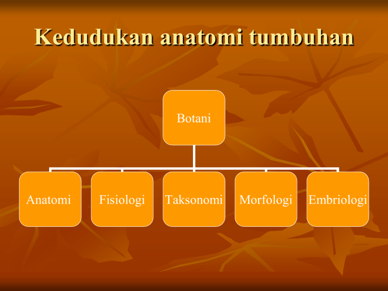
Kedudukan anatomi tumbuhan Botani Anatomi Fisiologi Taksonomi Morfologi Embriologi Sitologi tumbuhan : ilmu yang mempelajari bentuk, susunan, sifat fisik dan kimia sel tumbuhan serta perkembangan dinding selnya Histologi : ilmu yang mempelajari sekelompok atau sekumpulan sel yang membentuk suatu jaringan, dimana sekelompok sel tersebut memiliki ciri yang serupa, baik bentuk, sifat, maupun fungsinya sam Organologi :ilmu yang mempelajari alat-alat pada tubuh tumbuhan yang tampak dari luar, yaitu akar, batang, daun, bunga, buah, biji Konsep sel tumbuhan CELL -The cell is the structural and functional unit of all known living organisms. It is the smallest unit of an organism that is classified as living, and is often called the building block of life. Some organisms, such as most bacteria, are unicellular (consist of a single cell). Other organisms, such as humans, are multicellular. -Eukaryotic cells are about 10 times the size of a typical prokaryote and can be as much as 1000 times greater in volume. The major difference between prokaryotes and eukaryotes is that eukaryotic cells contain membrane-bound compartments in which specific metabolic activities take place. Most important among these is the presence of a cell nucleus, a membrane-delineated compartment that houses the eukaryotic cell's DNA. It is this nucleus that gives the eukaryote its name, which means "true nucleus." Other differences include Berdasarkan ada tidaknya selubung/membran inti, serta membran yang mengikat berbagai macam organela, sel dibedakan menjadi 2 : Prokariota sel yang tidak mempunyai membran inti/ membran yang mengikat organela-organela, DNA terkonsentrasi pada daerah yang disebut nukleooid contoh : bakteri dan cyanobakteri Eukariota sel yang mempunyai struktur yang komplek, inti dan organel lainnya terbungkus oleh membran inti dan terdapat pada suatu larutan semi cair disebut sitosol contoh : tumbuhan, hewan Perbedaan utama sel hewan dan sel tumbuhan : ► Pada tumbuhan memiliki membran plasma dengan dinding sel yang kuat pada hewan hanya mempunyai membran plasma saja ► Pada tumbuhan dijumpai plastida pada hewan tidak ada ► Pada sel tumbuhan beberapa vakuola yang kecilkecil dapat bersatu menjadi vakuola yang besar pada sel hewan vakuola tetap kecil Animal Cell Plant Cell Cell wall none yes Plastids no yes Vacuole One or more small vacuoles One, large central vacuole taking up 90% of cell volume Shape round rectangular Glyoxysomes no Some plant cells have glyoxysomes Centrioles Always present Only present in lower plant forms Lysosomes Occur in cytoplasm Usually not evident Plasma Membrane Only cell membrane Cell membrane & cell wall Chloroplast Don’t have chloroplast Have chloroplast PLANT CELL ANIMAL CELL EARLY MICROSCOPES Zacharias Janssen - made 1st compound microscope a Dutch maker of reading glasses (late 1500’s) Leeuwenhoek made a simple microscope (mid 1600’s) magnified 270X Early microscope lenses made images larger but the image was not clear Leeuwenhoek's microscope A) a screw for adjusting the height of the object being examined B) a metal plate serving as the body C) a skewer to impale the object and rotate it D) the lens itself, which was spherical MODERN MICROSCOPES A microscope is simple or compound depending on how many lenses it contains A lens makes an enlarged image & directs light towards you eye • A simple microscope has one lens • Similar to a magnifying glass • Magnification is the change in apparent size produced by a microscope COMPOUND MICROSCOPE A compound microscope has multiple lenses (eyepiece & objective lenses) STEREOMICROSCOPE creates a 3D image TOTAL MAGNIFICATION Powers of the eyepiece (10X) multiplied by objective lenses determine total magnification. ELECTRON MICROSCOPES More powerful; some can magnify up to 1,000,000X Use a magnetic field in a vacuum to bend beams of electrons Images must be photographed or produced electronically Scanning Electron Microscope (SEM) Electron microscope image of a spider produces realistic 3D image only the surface of specimen can be observed Electron microscope image of a fly foot Transmission Electron Microscope (TEM) produces 2D image of thinly sliced specimen detailed cell parts (only inside a cell) can be observed CELL THEORY • A theory resulting from many scientists’ observations & conclusions CELL THEORY 1. The basic unit of life is the cell. (Hooke) In 1665, an English scientist named Robert Hooke made an improved microscope and viewed thin slices of cork viewing plant cell walls Hooke named what he saw "cells" CELL THEORY 2. All living things are made of 1 or more cells. Matthias Schleiden (botanist studying plants) Theodore Schwann (zoologist studying animals) stated that all living things were made of cells Schwann Schleiden CELL THEORY 3. All cells divide & come from old cells. (Virchow) Virchow
