Uploaded by
faayzadudut
Intensive Care Nursery Staff Manual: Inborn Errors of Metabolism
advertisement
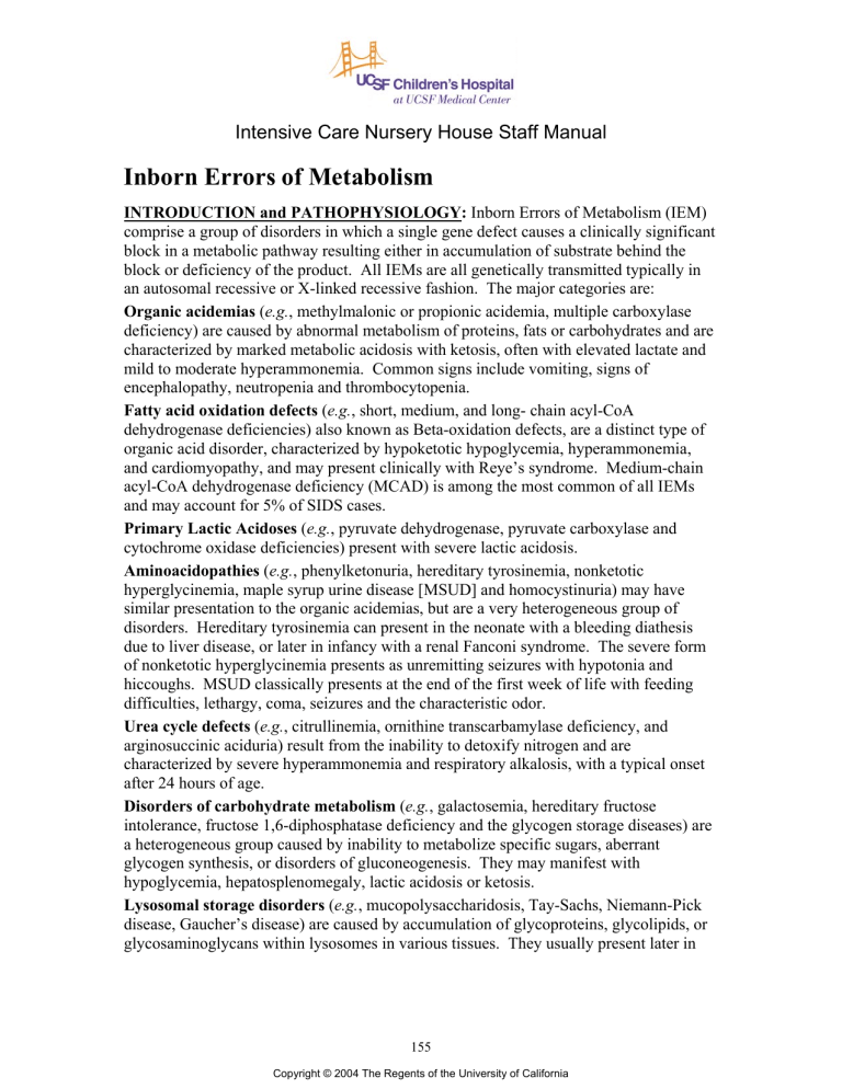
Intensive Care Nursery House Staff Manual Inborn Errors of Metabolism INTRODUCTION and PATHOPHYSIOLOGY: Inborn Errors of Metabolism (IEM) comprise a group of disorders in which a single gene defect causes a clinically significant block in a metabolic pathway resulting either in accumulation of substrate behind the block or deficiency of the product. All IEMs are all genetically transmitted typically in an autosomal recessive or X-linked recessive fashion. The major categories are: Organic acidemias (e.g., methylmalonic or propionic acidemia, multiple carboxylase deficiency) are caused by abnormal metabolism of proteins, fats or carbohydrates and are characterized by marked metabolic acidosis with ketosis, often with elevated lactate and mild to moderate hyperammonemia. Common signs include vomiting, signs of encephalopathy, neutropenia and thrombocytopenia. Fatty acid oxidation defects (e.g., short, medium, and long- chain acyl-CoA dehydrogenase deficiencies) also known as Beta-oxidation defects, are a distinct type of organic acid disorder, characterized by hypoketotic hypoglycemia, hyperammonemia, and cardiomyopathy, and may present clinically with Reye’s syndrome. Medium-chain acyl-CoA dehydrogenase deficiency (MCAD) is among the most common of all IEMs and may account for 5% of SIDS cases. Primary Lactic Acidoses (e.g., pyruvate dehydrogenase, pyruvate carboxylase and cytochrome oxidase deficiencies) present with severe lactic acidosis. Aminoacidopathies (e.g., phenylketonuria, hereditary tyrosinemia, nonketotic hyperglycinemia, maple syrup urine disease [MSUD] and homocystinuria) may have similar presentation to the organic acidemias, but are a very heterogeneous group of disorders. Hereditary tyrosinemia can present in the neonate with a bleeding diathesis due to liver disease, or later in infancy with a renal Fanconi syndrome. The severe form of nonketotic hyperglycinemia presents as unremitting seizures with hypotonia and hiccoughs. MSUD classically presents at the end of the first week of life with feeding difficulties, lethargy, coma, seizures and the characteristic odor. Urea cycle defects (e.g., citrullinemia, ornithine transcarbamylase deficiency, and arginosuccinic aciduria) result from the inability to detoxify nitrogen and are characterized by severe hyperammonemia and respiratory alkalosis, with a typical onset after 24 hours of age. Disorders of carbohydrate metabolism (e.g., galactosemia, hereditary fructose intolerance, fructose 1,6-diphosphatase deficiency and the glycogen storage diseases) are a heterogeneous group caused by inability to metabolize specific sugars, aberrant glycogen synthesis, or disorders of gluconeogenesis. They may manifest with hypoglycemia, hepatosplenomegaly, lactic acidosis or ketosis. Lysosomal storage disorders (e.g., mucopolysaccharidosis, Tay-Sachs, Niemann-Pick disease, Gaucher’s disease) are caused by accumulation of glycoproteins, glycolipids, or glycosaminoglycans within lysosomes in various tissues. They usually present later in 155 Copyright © 2004 The Regents of the University of California Inborn Errors of Metabolism infancy, not with a specific laboratory abnormality, but with organomegaly, facial coarseness and neurodegeneration and show a progressively degenerative course. Peroxisomal disorders (e.g., Zellweger syndrome and neonatal adrenoleukodystrophy) result from failure of the peroxisomal enzymes. They may present with features similar to the lysosomal storage disorders. Common features of Zellweger syndrome include large fontanel, organomegaly, Down-like facies, seizures and chondrodysplasia punctata. Others include disordered steroidogenesis (congenital adrenal hyperplasia or SmithLemli-Opitz), disorders of metal metabolism (Menkes syndrome, neonatal hemochromatosis). Transient hyperammonemia of the newborn is more prevalent in slightly premature infants receiving mechanical ventilation; onset is usually within the first 24 hours of life. The ammonia level may be markedly elevated and dialysis may be necessary. The cause is unknown and, if the newborn survives, there is no further evidence of impaired ammonia metabolism. CLINICAL FINDINGS suggestive of an IEM include: -History of consanguinity, mental retardation, or SIDS; symptom onset with institution of feedings or formula change; history of growth disturbances, lethargy, recurrent emesis, poor feeding, rashes, seizures, hiccoughs, apnea, tachypnea. -Physical findings: tachypnea, apnea, lethargy, hypertonicity, hypotonicity, hepatosplenomegaly, ambiguous genitalia, jaundice, dysmorphic or coarse facial features, rashes or patchy hypopigmentation, ocular findings (cataracts, lens dislocation or pigmentary retinopathy), intracranial hemorrhage, unusual odors. -Laboratory findings: metabolic acidosis with increased anion gap, primary respiratory alkalosis, hyperammonemia, hypoglycemia, ketosis or ketonuria, low BUN, hyperbilirubinemia, lactic acidosis, high lactate/pyruvate ratio, non-glucose-reducing substances in urine, elevated liver function tests including PT and PTT, neutropenia and thrombocytopenia. INITIAL APPROACH: -Rule out non-metabolic causes of symptoms such as infection or asphyxia. -Obtain consult from Genetic/Metabolic Service -Laboratory assessment prior to therapy: • Blood: glucose, newborn screen, CBC with differential, platelets, pH and PaCO2, electrolytes for anion gap, liver function tests, total and direct bilirubin, PT, PTT, uric acid, ammonia. Other studies may be indicated as described in algorithm below. Blood ammonia, lactate and pyruvate should be collected without a tourniquet, kept on ice and analyzed immediately. • Urine: color, odor, pH, glucose, ketones, reducing substances (positive for galactosemia, fructose intolerance, tyrosinemia and others), ferric chloride reaction (positive for MSUD, PKU and others) and DNPH reaction (screens for alpha-keto acids). Amino and organic acids as described in algorithm below. • CSF: glycine (for nonketotic hyperglycinemia), lactate, pyruvate if appropriate. -Figures 1 and 2 are adapted from Burton BK: Inborn errors of metabolism in infancy: A guide to diagnosis. Pediatrics 102:E69, 1998. These flow charts are guides to the differential diagnosis of hyperammonemia (Figure 1) and metabolic acidosis (Figure 2 ) in newborns. Another helpful algorithm is in Rudolph’s Pediatrics, 20th ed., P. 292. Not all inborn errors of metabolism will present with acidosis, hyperammonemia, 156 Inborn Errors of Metabolism or hypoglycemia. Neurological signs (e.g., seizures, obtundation) may be the predominant feature in several IEMs (e.g., nonketotic hyperglycinemia, molybdenum cofactor deficiency, peroxisomal disorders). NEONATAL HYPERAMMONEMIA Symptoms in 1st 24h Preterm Symptoms after age 24h Transient Hyperammonemia of Newborn No acidosis Acidosis Full Term INBORN ERROR OF METABOLISM (i.e., organic acidemia or PC deficiency) UREA CYCLE DEFECTS ORGANIC ACIDEMIAS PLASMA AMINO ACIDS Absent Citrulline Citrulline moderately elevated, ASA present Urine Orotic Acid Low CPS deficiency Arginosuccinic aciduria Elevated Citrulline markedly Elevated, no ASA Citrullinemia OTC deficiency Figure 1. Flow chart for differential diagnosis of hyperammonemia. (ASA, arginosuccinic acid; CPS, carbamyl phosphate synthetase; OTC, ornithine transcarbamylase; PC, pyruvate carboxylase). Chart is adapted from Burton BK: Pediatrics 102: E69, 1998. FURTHER MANAGEMENT: Although treatment differs for each specific disorder, each should be managed by considering the potential disease category and by following the following steps, along with consultation with a metabolic specialist. -Hydration/nutrition/acid-base management: Rehydrate infant. Stop all oral intake to eliminate protein, galactose and fructose. Provide calories with IV glucose at 8-10 mg/kg/min (even if insulin is required to keep the blood glucose level normal). Give IV lipids only after ruling out a primary or secondary fatty acid oxidation defect. Withhold all protein for 48 to 72 hours, while the patient is acutely ill, and until an aminoacidopathy, organic aciduria or urea cycle defect has been excluded. Special special enteral formulas and parenteral amino acid solutions are available for many disorders. Treat significant acidosis (pH<7.22) with a continuous infusion of NaHCO3. 157 Inborn Errors of Metabolism -Elimination of toxic metabolites: Treatment of hyperammonemia is urgent. The severity of neurological impairment in infants with urea cycle defects depends upon the duration of the hyperammonemic coma. For severe hyperammonemia, hemodialysis is indicated. Dialysis may also be indicated for intractable anion gap metabolic acidosis. METABOLIC ACIDOSIS WITH INCREASED ANION GAP Normal lactate Abnormal organic acids Elevated lactate Normal organic acids Abnormal organic acids ORGANIC ACIDEMIA Elevated pyruvate; normal L:P ratio Dicarboxylic aciduria FATTY ACID OXIDATION DEFECTS Hypoglycemia Normal or low pyruvate; elevated L:P ratio RESPIRATORY CHAIN DEFECTS; PYRUVATE CARBOXYLASE DEFICIENCY No hypoglycemia METHYLMALONIC ACIDEMIA;PROPIONIC ACIDEMIA; MULTIPLE CARBOXYLASE DEFICIENCY; OTHERS GSD TYPE 1; FRUCTOSE 1,6-DP DEFICIENCY; PEP CARBOXYKINASE DEFICIENCY PYRUVATE DEHYDROGENASE DEFICIENCY; PYRUVATE CARBOXYLASE DEFICIENCY Figure 2. Flowchart for evaluation of metabolic acidosis in the young infant. (Fructose-1,6-DP, fructose-1,6-diphosphatase; GSD, glycogen storage disease; L:P ratio, lactate to pyruvate ratio) Chart is adapted from Burton BK: Pediatrics 102: E69, 1998. ________________________________________________________________________ -Treatment of coexisting/precipitating factors (e.g., infection, thrombocytopenia). -Cofactor replacement: Certain enzyme deficiencies are vitamin-responsive, including: • The vitamin-responsive form of propionic acidemia, beta-methylcrotonyl deficiency, holocarboxylase synthetase deficiency, and biotinidase deficiency: biotin (5 mg daily, oral or parenteral). • Vitamin-responsive methylmalonic acidemia: Vitamin B12 (1 mg daily, IM). • Vitamin-responsive maple syrup urine disease: thiamine (50 mg daily, oral). 158 Inborn Errors of Metabolism It is important to make a specific diagnosis, even in a dying child, to help parents understand what happened and to provide information that might affect future reproductive planning. -If an autopsy is not permitted, request consent for pre-mortem or immediately postmortem specimens. -Blood should be centrifuged and the plasma should be frozen. -Urine and spinal fluid should be refrigerated. -A sterile skin biopsy (used for fibroblast culture) can be performed within 1-2 hours after death. Alcohol should be used to clean the skin; do not use betadine, as it will inhibit cell growth. Place skin sample in sterile saline at room temperature and send to the cytogenetics lab for culture and processing. -Other tissue samples (e.g., liver, skeletal muscle, cardiac muscle, brain, kidney) may be useful depending on the disorder. -If there are dysmorphic features, consider a full skeletal radiological series. OUTCOME: At present, for most IEMs, prognosis for survival or normal neurological outcome is guarded, despite appropriate and aggressive therapy. It is likely that the outcomes will improve with (a) presymptomatic diagnosis (i.e., by prenatal detection or expanded neonatal screening), (b) identification of genes and other factors which impact on phenotype, response to treatment, and outcome, and (c) alternative novel approaches to therapy. 159
