kata pengantar
advertisement
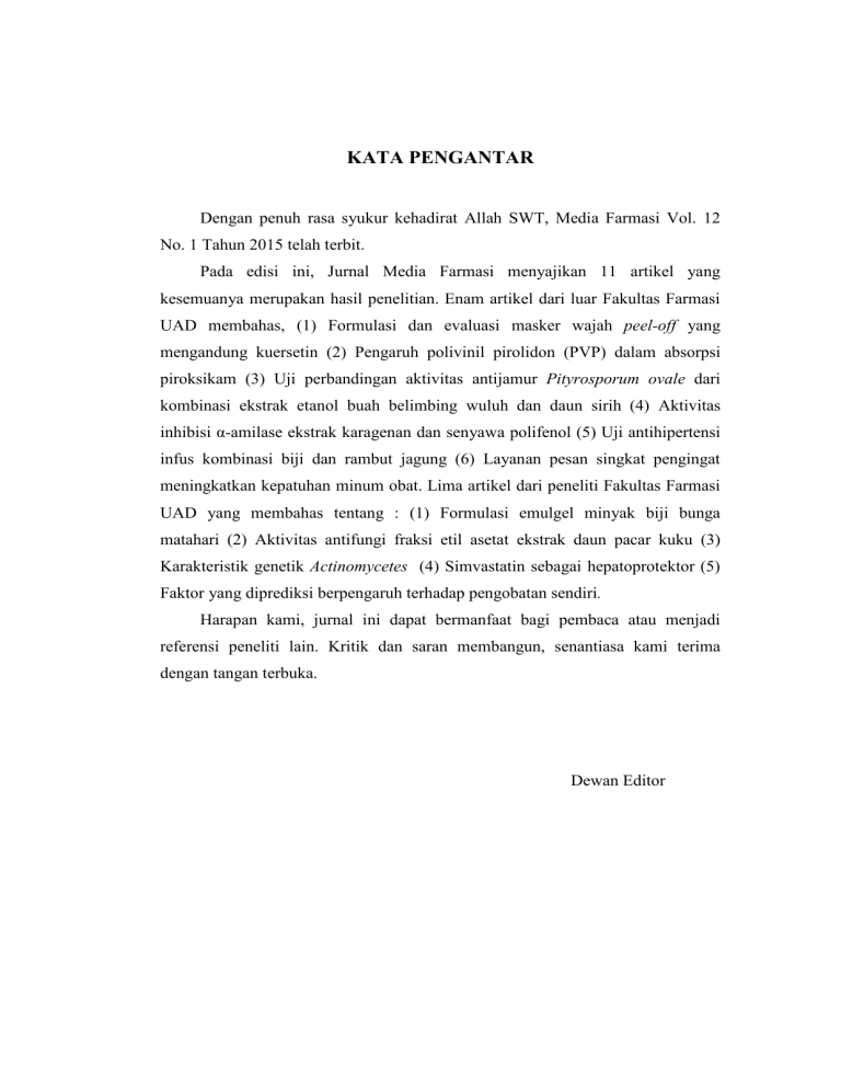
KATA PENGANTAR Dengan penuh rasa syukur kehadirat Allah SWT, Media Farmasi Vol. 12 No. 1 Tahun 2015 telah terbit. Pada edisi ini, Jurnal Media Farmasi menyajikan 11 artikel yang kesemuanya merupakan hasil penelitian. Enam artikel dari luar Fakultas Farmasi UAD membahas, (1) Formulasi dan evaluasi masker wajah peel-off yang mengandung kuersetin (2) Pengaruh polivinil pirolidon (PVP) dalam absorpsi piroksikam (3) Uji perbandingan aktivitas antijamur Pityrosporum ovale dari kombinasi ekstrak etanol buah belimbing wuluh dan daun sirih (4) Aktivitas inhibisi α-amilase ekstrak karagenan dan senyawa polifenol (5) Uji antihipertensi infus kombinasi biji dan rambut jagung (6) Layanan pesan singkat pengingat meningkatkan kepatuhan minum obat. Lima artikel dari peneliti Fakultas Farmasi UAD yang membahas tentang : (1) Formulasi emulgel minyak biji bunga matahari (2) Aktivitas antifungi fraksi etil asetat ekstrak daun pacar kuku (3) Karakteristik genetik Actinomycetes (4) Simvastatin sebagai hepatoprotektor (5) Faktor yang diprediksi berpengaruh terhadap pengobatan sendiri. Harapan kami, jurnal ini dapat bermanfaat bagi pembaca atau menjadi referensi peneliti lain. Kritik dan saran membangun, senantiasa kami terima dengan tangan terbuka. Dewan Editor Genetic Characteristics of Actinomycetes Melati dan Nanik 57 GENETIC CHARACTERISTICS OF ACTINOMYCETES ISOLATES (GST, KP, KP11, KP16, T24, T37 CODE) BY RFLP OF NRPS GENES Melati Aprilliana Ramadhani, Nanik Sulistyani Faculty of Pharmacy, Ahmad Dahlan University, Yogyakarta, Indonesia Email : [email protected] ABSTRACT Actinomycetes is the one of antibiotic producing microorganisms. Actinomycetes isolates with GST, KP, KP11, KP16, T24, T37 have been isolated from soil. This study aims to determine the diversity of actinomycetes isolates by RFLP (Restriction Fragment Length Polymorphism) of NRPS (Non Ribosomal Peptide Synthetase) genes from GST, KP, KP11, KP16, T24, and T37 isolates. This study is divided into several steps of the DNA isolation from isolate GST, KP16, T24, T37, KP, KP11, PCR of the 16S rRNA and NRPS genes and cutting the NRPS genes fragmens with HaeIII enzyme to determine the diversity of isolates. Results of DNA isolate, PCR of 16S rRNA genes and NRPS genes, and RFLP of NRPS genes performed by agarose gel electrophoresis. The diversity of the isolates were analyzed by multivariate analysis UPGMA with MVSP 2.0 applications. The results showed the diversity of 6 actinomycetes isolates are T37 and GST have 64 % similarity, KP11 and KP16 have 96 % similarity, KP has 76 % similarity with KP11 and KP16, while T24 has 60 % similarity with KP and 12 % similarity with T37 and GST. Based on RFLP analysis of the NRPS genes, among of 6 isolates can divided into 4 groups are T37 and GST isolates as group 1, KP11 and KP16 isolates as group 2, KP isolate as group 3, and T24 isolate as group 4. Keyword : Actinomycetes, diversity, 16S rRNA genes, NRPS genes, RFLP microorganisms, among others by INTRODUCTION NRPS (Nonribosomal peptide synthetase) is biosynthetic systems involved in the synthesis of a large number of important biologically active compounds produced by actinomycetes (Sacido et al., 2005). NRPS is multi-enzymatic, multidomain megasynthases involved in the biosynthesis of nonribosomal peptides. These secondary 58 metabolites exhibit a remarkable array of biological activity and many Media Farmasi Vol 12 No.1 Maret 2015 : 57-65 Genetic Characteristics of Actinomycetes Melati dan Nanik 59 of them are clinically valuable anti- polymorphic sequences, is a simple, microbial, anti-fungal, anti-parasitic, reliable, anti-tumor and immunosuppressive inexpensive method with minimum agents. Nonribosomal peptides are equipment requirements. RFLP is biosynthesized based on the creation or deletion of condensation by of sequential amino acid relatively NRPS modules contain the and recognition site of a restriction endonuclease monomers (Ansari et al., 2004). fast, by nucleotide variations in the polymorphic site; the thus, digestion of the PCR product and containing the polymorphism with an thiolation steps involved in the appropriate restriction endonuclease recognition and condensation of the results in disparate electrophoretic substrate. patterns by polymorphism genotype activities corresponding condensation, to adenylation, Additional (heterocyclase, domains N-methylase, (Sharafi et al., 2012). and The principle of this method is reductase) are also present depending cutting gene become fragments with on the requirements for the substrate restriction enzyme. Differences size activation, of the fragments cutting result, epimerase, thioesterase, elongation, and modification (Sacido et al., 2005). In showed the natural analyzed. In the case, the RFLP of products screening programs have NRPS genes profile, then indicates concentrated an intense effort in the the discovery secondary metabolites are resulting previous of metabolites decades, biologically produced active by actinomycetes (Sacido et al., 2005). of gene existence diversity of are differences (Faizal et al., 2008). The objective of this research Genetic diversity of NRPS was to observe the diversity of 6 genes isolates of actinomycetes can actinomycetes isolates with GST, be analyzed by the method of KP16, T24, T37, KP, and KP11 code Restriction Length based on the RFLP of NRPS genes RFLP profile. The results of the research amplified can be used to identify the diversity Polymorphism known as Fragment (RFLP). cleaved 60 Media Farmasi Vol 12 No.1 Maret 2015 : 57-65 and characterization of actinomycetes of 1-2 ml was taken, and then centrifuged with 5000 rpm actinomycetes isolates. 10 minutes. Pellet was washed with 200 µl of buffer TE. Mixed pellets METHODOLOGY The tools used in this research are: glass tools, PCR, electrophoresis, UV lamp. The materials used are the SNB media, agarose 1%, agarose 2%, Etidium bromide, DNA Ladder (Marker), A7R and A3F primary, F27 and R1492 primary, buffer R, enzyme BsuRI. Culture preparation has been done by filling 20 ml starter inside an containing 200 ml medium SNB already sterilized. The media containing 20 ml starter, was incubated for 5 days by stirer with magnetic stirring. The preparation of this culture has been done at LAF room to minimize the occurrence of (Sulistyani et al, 2013). DNA isolation method based Farida modification. et hours, after 1 hour add with 50 µl NaCl 5M and inversion every 30 minutes, centrifuged at 13000 rpm 10 minutes, water phase was moved to a new tube. DNA obtained from the water phase, add with cold was centrifuged at 13000 rpm 5 minutes, supernatan discarded. DNA pellet was cleaned with 50 µl ethanol 70%, centrifuged 13000 rpm 10 minutes. Resuspension pellet with buffer TE 50 µl. Electrophoresis DNA sample of 5 μl mixed with 1 µL DNA loading, resuspension and put in the well of gel agarosa 1% and connected the electrode to the power supply with DNA Isolation on 50µl SDS, incubated at 65OC for 2 stored overnight at 20°C. Sample Culture Preparation contamination for 60 minutes and then add with isopropanol as much as 50µl, and DNA Isolation erlenmeyer with 50 µl lisosim, incubated at 37OC al. The (2007) isolates with of 100 volts voltage for 30 minutes, then electrophoresis. The result of electrophoresis was seen on UV Genetic Characteristics of Actinomycetes Melati dan Nanik 61 transluminator and observed the 72°C for 4 minutes, and the final ribbons of DNA visualisasion. extension at 72°C for 5 minutes. The Fragmen Amplification of 16S rRNA program is carried out as many as 35 and NRPS DNA genes cycles, then stop the process by DNA fragments of 16S rRNA setting the temperature at 4°C genes was amplified with PCR (Sacido et al, 2005). Composition of methods use F27 and R1492 primer, materials for PCR of NRPS genes are while NRPS use A3F and A7R KIF primary (2µl), M6R primary primer. PCR for 16S rRNA genes (2µl), was performed with the following nuclease free water (11µl), and green program: initial denaturation at 96°C PCR (25µl). All of the ingredients for 3 minutes, then 94°C for 1 filled an eppendrof, mixed and add minute, the process of anneling at mineral oil 20 μl so that evaporation 53°C for 1 minute, the extension at does not occur. After PCR was done, 72°C for 5 minutes, and the final mineral oil and DNA was separated extension at 72 ° C for 5 minutes. then entered a new eppendorf and The program is carried out as many stored at 20°C. PCR products was as 30 cycles, then stop the process by visualized using electrophoresis gel setting agarosa 1% by putting PCR product the temperature at 4°C DNA templete (10µl), (Sulistyani, 2013). Composition of into the well as much as 5 μl. materials for PCR of 16S rRNA Analysis RFLP of NRPS genes genes are F27 primary (0,5µl), PCR product is cutting by DNA restriction BsuRI (HaeIII) enzym, templete (10µl), nuclease free water then electroforesis with agarose gel (14µl), and green PCR (25µl). 2%. Composition of materials for R1492 primary (0,5µl), PCR for NRPS genes was perfomed with the following RFLP of NRPS genes are PCR NRPS product (7.5µl), aqua (5µl), program: initial denaturation at 96°C enzim BsuRI (1u/µl) (2µl), mixed the for 5 minutes, then 95°C for 30 composition, incubated overnight at seconds, the process of anneling at 37OC. Enzyme BsuRI (1u/µl) made 58°C for 2 minutes, the extension at 62 Media Farmasi Vol 12 No.1 Maret 2015 : 57-65 by mixed 2 µl of enzyme HaeIII very clear fragment, while the other (BsuRI) with buffer R 18 µl. isolates looks very thin fragment, but can be proceed to the next steps. RESULT AND DISCUSSION Spectrophotometric UV-Vis Spectrophotometry DNA Isolation analysis DNA isolation is the first step from various DNA analysis technology. DNA can be found in the core or on chromosome organelles either. DNA isolation was broken the cell wall and nucleus membrane, followed by separation of DNA from other cell components. Electrophoresis have been done after pure DNA obtained to identify presence of DNA. Isolation of DNA results can be seen in Figure 1. method based is an on the measurement of UV ray absorption monokromatis by a strip of colored solution at a spesific wavelength by using a prism monokromator or diffraction grating with a fototube detector (Sudarmadji, 1996). DNA concentration can be determined using spectrophotometer UV-rays at the wavelength of 260 nm. The amount of radiation absorbed by solution (Absorban) is directly proportional to the concentration of DNA. Absorban comparison (ratio) at the wavelength of 260 and 280 nm can indicated the purity of the isolate DNA. If the ratio less than 1,8 its means that DNA isolate contamined by protein and if the ratio more than Figure 1. Results of electrophoresis of DNA isolation of actinomycetes (Wells Order: GST, KP16, T24, T37, KP, KP11, Marker) 2,0 its means that DNA isolate Figure I shows that all isolates 2003). Results of Spectrophotometric contamined by RNA (Stephenson, of actinomycetes can be isolated with antibiotic a size more than 3000 bp. Although isolate DNA in table I. only KP16 and T24 isolates looks producing bacteria of Genetic Characteristics of Actinomycetes Melati dan Nanik 63 Table I. Measurement results OD at 260 nm and 280 nm No Sample OD at 260 nm OD at 280 nm Purity of DNA (OD 260nm/OD 280 nm) 1. KP16 0.0098 0.0065 1.51 2. GST 0.0081 0.0044 1.84 3. T24 0.0111 0.0015 7.40 4. T37 0.0054 0.0017 3.18 5. KP 0.0289 0.0232 1.25 6. KP11 0.0034 0.0039 0.87 Figure 2. Results of Electrophoresis PCR DNA of 16S rRNA genes (wells order: Marker, GST, KP16, T24, T37, KP, KP11) Purity results of DNA measured isolation by PCR DNA 16S rRNA GENES UV PCR DNA 16S rRNA genes spectrophotometry at 260 nm and was done for ensure that DNA from 280 nm. Based on the results KP16, all KP, and KP11 have the results with amplificated. 16S rRNA genes used a ratio less than 1,8 its means the as indicators of the amplification possibility contamination by protein, process. Results of electrophoresis and a ratio more than 2,0 possibility PCR DNA of 16S rRNA genes in contamination by RNA, but tried to Figure 2. continued on PCR DNA 16S rRNA gene. actinomycetes isolates can Results of the PCR as showed in Figure 2 that the DNA of actinomycetes isolates are amplicable because all of 16S rRNA 64 Media Farmasi Vol 12 No.1 Maret 2015 : 57-65 genes was detected and measuring primary use for amplification of 1200 bp, except PCR product of NRPS genes are A3F (forward T24, T24 fragment looks relegated, primer) and A7R (reverse primer). however can be continued to the This primer will stick in the process PCR of NRPS genes. adenylation domain on NRPS genes PCR DNA NRPS GENES and produce PCR product that are NRPS genes was involved in have a measurement of 700-800 bp production of secondary metabolites (Sacido et al., 2005). PCR results that are in Figure 3. are important pharmaceutical industry. in the NRPS Based on the results of genes detection has been performed electrophoresis, all of PCR product to explore the biosynthetic potential of NRPS genes have a size of 700 across bp. all major actinomycetes liniages (Sacido et al., 2005). The Figure 3. Results of Electrophoresis of PCR DNA of NRPS genes (wells order : GST, KP16, T24, T37, KP, KP11, Marker) Figure 4. Results of Electrophoresis of RFLP NRPS genes (wells order: GST, KP16, T24, T37, KP, KP11, RFLP Marker) Genetic Characteristics of Actinomycetes RFLP of NRPS genes Melati dan Nanik 65 HaeIII enzyme will be showed the RFLP method was used to different profiles in different know the cutting results of DNA actinomycetes isolates are shown in fragments profile of NRPS genes of figure 4 and table II. actinomycetes isolates by HaeIII Diversity profiles can be enzyme. HaeIII enzyme cutting the observed from size of fragments by DNA of NRPS genes amplification enzim HaeIII cutting results, then th results in areas which rich in GC size of fragments input to UPGMA base pairs. This enzyme has the multivariate analysis with MVSP identification of four bases are cut in 2.0 areas that have a base sequence of dondrogram. The results in figure 5. GGCC (Dharmaraj, 2010). Therefore, the results of cutting application to array the Based on the image can be grouped that T37 and GST have 64 Table II. Restriction Fragments Size Results in each PCR of NRPS genes Product Isolat Code GST KP16 T24 T37 KP KP11 Size (bp) 288, 209, 102 933, 724 933, 288, 102 288 933, 724, 389, 363, 209, 102 933, 724, 102 Figure 5. UPGMA Analysis Multivariate Results of the Cutting NRPS Genes By Enzyme HaeIII 66 Uji Perbandingan Aktivitas Siti Sakinah, dkk % similarity, KP11 and KP16 have 96 % similarity, KP has 76 % similarity with KP11 and KP16, while T24 has 60 % similarity with KP and 12 % similarity with T37 and GST. CONCLUSION Based on RFLP analysis of the NRPS genes, actinomycetes among isolates of 6 can be classified into 4 groups are T37 and GST isolates as group 1, KP11 and KP16 isolates as group 2, KP isolate as group 3, and T24 isolate as group 4. REFERENCES Ansari, M.Z; Yadav, G; Gokhale, R.S; and Mohanti, D., 2004, NRPS-PKS: a knowledge-besed resource for analysis of NRPS/PKS megasynthases, Nucleic Acids Research, Vol. 32: W405-W413. Dharmaraj, 2010, Marine Streptomyces as a novel source of bioactive substance, World J Microbiol Biotechnol, 26:21232139. Faizal et al, 2008, Polymorphism Analysis of Polyketide Synthase Gene from Actinomycetes Genome DNA of Taman Nasional Gunung Halimun Soil by Using Metagenome Method, Journal of Biotechnology Research in Tropical Region, Vol 1, Jakarta. Farida,, Widada, Meiyanto, 2007, Combination Methods for Screening Marine Actinomycetes Producing Potential Compounds as Anticancer, Indonesian Journal of Biotechnology, Vol. 12. No. 2, pp. 988-997. Sacido, A., Genilloud, O., 2004, New PCR Primers for the Screening of NRPS and PKS-I Systems in Actinomycetes: Detection and Distribution of These Biosynthetic Gene Sequences in Major Taxonomic Groups, Microbial Ecology, Spain. Sharafi et al, 2012, Development and Validation of a Simple, Rapid and Inexpensive PCR RFLP Method for Genotyping of Common IL28B Polymorphisms: A Useful Pharmacogenetic Tool for Prediction of Hepatitis C Treatment Response, Hepatitis Monthly, 12(3):190-195 Sudarmadji, S., Suhardi., 1996, Analisis Bahan Makanan dan Pertanian, Liberty, Yogyakarta. Sulistyani, N., 2013, Keragaman Isolat Actinomycetes Berdasarkan Analisis RFLP Terhadap Gen NRPS, Jurnal Ilmiah Kefarmasian, Vol. 3, No. 1, 2013:81-94. Stephenson, F.H., 2003, Calculation in Molecular Biology and Biotechnology, A Guide to Mathematics in The Laboratory, Academic Press, California, USA.

